9H80 | pdb_00009h80

Structure of the outer membrane exopolysaccharide transporter PelBC


Experimental Data Snapshot

  • Method: ELECTRON MICROSCOPY
  • Resolution: 2.50 Å
  • Aggregation State: PARTICLE 
  • Reconstruction Method: SINGLE PARTICLE 

wwPDB Validation   3D Report Full Report


This is version 1.1 of the entry. See complete history


Literature

Assembly and the gating mechanism of the Pel exopolysaccharide export complex PelBC of Pseudomonas aeruginosa.

Benedens, M.Rosales-Hernandez, C.Straathof, S.A.P.Loschwitz, J.Berninghausen, O.Maglia, G.Beckmann, R.Kedrov, A.

(2025) Nat Commun 16: 5249-5249

  • DOI: https://doi.org/10.1038/s41467-025-60605-8
  • Primary Citation of Related Structures:  
    9H80

  • PubMed Abstract: 

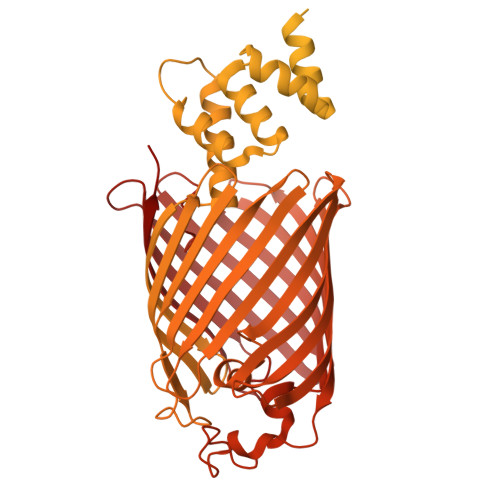
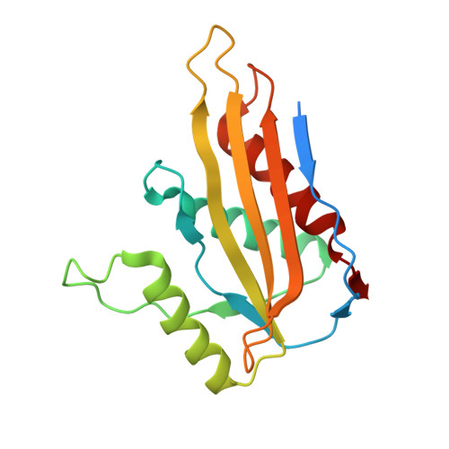
    The pathogen Pseudomonas aeruginosa enhances its virulence and antibiotic resistance upon formation of durable biofilms. The exopolysaccharides Pel, Psl and alginate essentially contribute to the biofilm matrix, but their secretion mechanisms are barely understood. Here, we reveal the architecture of the outer membrane complex PelBC for Pel export, where the essential periplasmic ring of twelve lipoproteins PelC is mounted on top of the nanodisc-embedded β-barrel PelB. The PelC assembly is stabilized by electrostatic contacts with the periplasmic rim of PelB and via the membrane-anchored acyl chains. The negatively charged interior of the PelB β-barrel forms a route for the cationic Pel exopolysaccharide. The β-barrel is sealed at the extracellular side, but molecular dynamic simulations suggest that the short loop Plug-S is sufficiently flexible to open a tunnel for the exopolysaccharide transport. This gating model is corroborated by single-channel conductivity measurements, where a deletion of Plug-S renders a constitutively open β-barrel. Our structural and functional analysis offers a comprehensive view on this pathogenicity-relevant complex and suggests the route taken by the exopolysaccharide at the final secretion step.


  • Organizational Affiliation
    • Synthetic Membrane Systems, Institute of Biochemistry, Heinrich Heine University Düsseldorf, Düsseldorf, Germany.

Macromolecules
Find similar proteins by:  (by identity cutoff)  |  3D Structure
Entity ID: 1
MoleculeChains Sequence LengthOrganismDetailsImage
PelBA [auth M]1,193Pseudomonas aeruginosaMutation(s): 0 
Gene Names: pelBPA3063
UniProt
Find proteins for Q9HZE5 (Pseudomonas aeruginosa (strain ATCC 15692 / DSM 22644 / CIP 104116 / JCM 14847 / LMG 12228 / 1C / PRS 101 / PAO1))
Explore Q9HZE5 
Go to UniProtKB:  Q9HZE5
Entity Groups  
Sequence Clusters30% Identity50% Identity70% Identity90% Identity95% Identity100% Identity
UniProt GroupQ9HZE5
Sequence Annotations
Expand
  • Reference Sequence
Find similar proteins by:  (by identity cutoff)  |  3D Structure
Entity ID: 2
MoleculeChains Sequence LengthOrganismDetailsImage
PelC172Pseudomonas aeruginosaMutation(s): 0 
Gene Names: pelCPA3062
UniProt
Find proteins for Q9HZE6 (Pseudomonas aeruginosa (strain ATCC 15692 / DSM 22644 / CIP 104116 / JCM 14847 / LMG 12228 / 1C / PRS 101 / PAO1))
Explore Q9HZE6 
Go to UniProtKB:  Q9HZE6
Entity Groups  
Sequence Clusters30% Identity50% Identity70% Identity90% Identity95% Identity100% Identity
UniProt GroupQ9HZE6
Sequence Annotations
Expand
  • Reference Sequence
Small Molecules
Ligands 1 Unique
IDChains Name / Formula / InChI Key2D Diagram3D Interactions
PTY (Subject of Investigation/LOI)
Query on PTY

Download Ideal Coordinates CCD File 
AA [auth H]
BA [auth I]
CA [auth I]
DA [auth I]
EA [auth J]
AA [auth H],
BA [auth I],
CA [auth I],
DA [auth I],
EA [auth J],
FA [auth J],
GA [auth K],
HA [auth K],
IA [auth L],
JA [auth L],
KA [auth A],
LA [auth A],
MA [auth B],
N [auth M],
NA [auth B],
O [auth M],
P [auth C],
Q [auth C],
R [auth D],
S [auth D],
T [auth E],
U [auth E],
V [auth F],
W [auth F],
X [auth G],
Y [auth G],
Z [auth H]
PHOSPHATIDYLETHANOLAMINE
C40 H80 N O8 P
NJGIRBISCGPRPF-KXQOOQHDSA-N
Experimental Data & Validation

Experimental Data

  • Method: ELECTRON MICROSCOPY
  • Resolution: 2.50 Å
  • Aggregation State: PARTICLE 
  • Reconstruction Method: SINGLE PARTICLE 
EM Software:
TaskSoftware PackageVersion
MODEL REFINEMENTPHENIX1.20.1_4487:

Structure Validation

View Full Validation Report


Entry History & Funding Information

Deposition Data


Funding OrganizationLocationGrant Number
German Research Foundation (DFG)Germany--

Revision History  (Full details and data files)

  • Version 1.0: 2025-06-11
    Type: Initial release
  • Version 1.1: 2025-06-18
    Changes: Data collection, Database references