


A cooperative binding mechanism steers the recruitment of peripheral subunits in the E. coli pyruvate dehydrogenase complex
Bothe, S.N., Racunica, D., Zajec Hudnik, T., Zdanowicz, R., Bothe, A., Giese, C., Glockshuber, R.To be published.
Experimental Data Snapshot
Starting Model: experimental
View more details
wwPDB Validation 3D Report Full Report

Entity ID: 1 | |||||
|---|---|---|---|---|---|
| Molecule | Chains | Sequence Length | Organism | Details | Image |
| Pyruvate dehydrogenase E1 component | A [auth C], B | 49 | Escherichia coli | Mutation(s): 0 Gene Names: aceE, b0114, JW0110 EC: 1.2.4.1 | 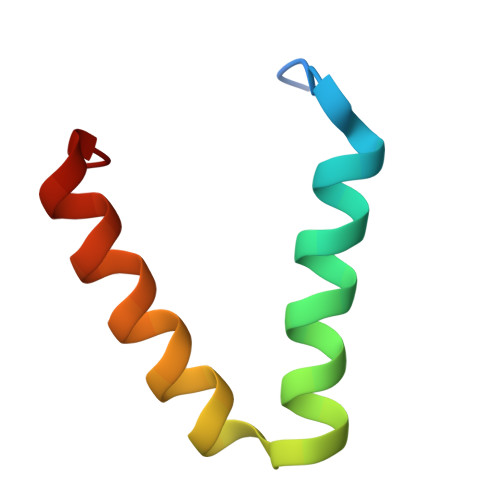 |
UniProt | |||||
Entity Groups | |||||
| Sequence Clusters | 30% Identity50% Identity70% Identity90% Identity95% Identity100% Identity | ||||
| UniProt Group | P0AFG8 | ||||
Sequence AnnotationsExpand | |||||
Reference Sequence | |||||
Entity ID: 2 | |||||
|---|---|---|---|---|---|
| Molecule | Chains | Sequence Length | Organism | Details | Image |
| Dihydrolipoyllysine-residue acetyltransferase component of pyruvate dehydrogenase complex | C [auth A] | 66 | Escherichia coli | Mutation(s): 0 Gene Names: aceF, b0115, JW0111 EC: 2.3.1.12 | 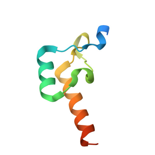 |
UniProt | |||||
Entity Groups | |||||
| Sequence Clusters | 30% Identity50% Identity70% Identity90% Identity95% Identity100% Identity | ||||
| UniProt Group | P06959 | ||||
Sequence AnnotationsExpand | |||||
Reference Sequence | |||||
| Length ( Å ) | Angle ( ˚ ) |
|---|---|
| a = 87.262 | α = 90 |
| b = 87.262 | β = 90 |
| c = 42.341 | γ = 90 |
| Software Name | Purpose |
|---|---|
| PHENIX | refinement |
| PHENIX | refinement |
| XDS | data reduction |
| Aimless | data scaling |
| PHASER | phasing |
| Funding Organization | Location | Grant Number |
|---|---|---|
| Swiss National Science Foundation | Switzerland | -- |


