


NMHase, dihydrouridine, 2.1A, CC_mask=0.7859
Toedtli, P., Rudolph, M.G.To be published.
Experimental Data Snapshot
wwPDB Validation 3D Report Full Report

Entity ID: 1 | |||||
|---|---|---|---|---|---|
| Molecule | Chains | Sequence Length | Organism | Details | Image |
| N-Methylhydantoinase | 1,288 | Glutamicibacter protophormiae | Mutation(s): 0 | 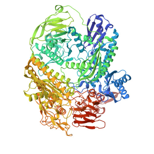 | |
UniProt | |||||
Entity Groups | |||||
| Sequence Clusters | 30% Identity50% Identity70% Identity90% Identity95% Identity100% Identity | ||||
| UniProt Group | M7MZT8 | ||||
Sequence AnnotationsExpand | |||||
Reference Sequence | |||||
| Ligands 2 Unique | |||||
|---|---|---|---|---|---|
| ID | Chains | Name / Formula / InChI Key | 2D Diagram | 3D Interactions | |
| DUC (Subject of Investigation/LOI) Download:Ideal Coordinates CCD File | D [auth A], F [auth B] | DIHYDROPYRIMIDINE-2,4(1H,3H)-DIONE C4 H6 N2 O2 OIVLITBTBDPEFK-UHFFFAOYSA-N |  | ||
| CA Download:Ideal Coordinates CCD File | C [auth A], E [auth B] | CALCIUM ION Ca BHPQYMZQTOCNFJ-UHFFFAOYSA-N |  | ||
| Funding Organization | Location | Grant Number |
|---|---|---|
| F. Hoffmann-La Roche LTD | Switzerland | -- |


