9GYA | pdb_00009gya

Vitamin D Receptor in complex with Sila-e


Experimental Data Snapshot

  • Method: X-RAY DIFFRACTION
  • Resolution: 1.85 Å
  • R-Value Free: 
    0.232 (Depositor), 0.234 (DCC) 
  • R-Value Work: 
    0.188 (Depositor), 0.192 (DCC) 
  • R-Value Observed: 
    0.191 (Depositor) 

Starting Model: experimental
View more details

wwPDB Validation 3D Report Full Report

Validation slider image for 9GYA

Ligand Structure Quality Assessment 


This is version 1.1 of the entry. See complete history

Literature

First Sila-Vitamin D Analogues: Design, Synthesis, Structural Analysis and Biological Activity.

Loureiro, J.Seoane, S.Sampaio-Dias, I.E.Peluso-Iltis, C.Guiberteau, T.Brito, B.Gregorio, C.Perez-Fernandez, R.Rochel, N.Mourino, A.Rodriguez-Borges, J.E.

(2024) J Med Chem 67: 21505-21519

  • DOI: https://doi.org/10.1021/acs.jmedchem.4c02404
  • Primary Citation Related Structures: 
    9GY8, 9GYA, 9GYC, 9GYJ, 9GYK

  • PubMed Abstract: 

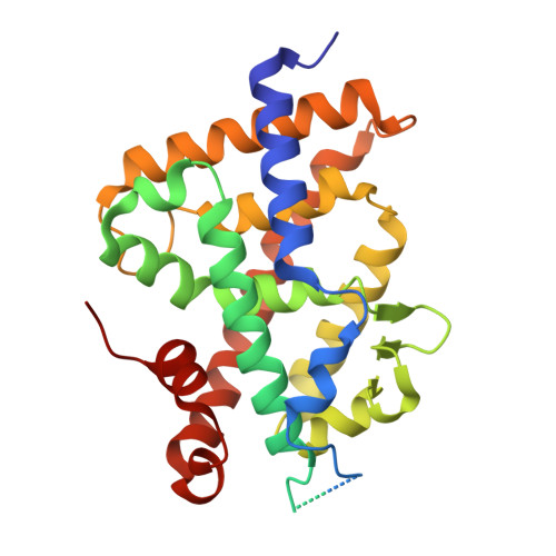
    The incorporation of silicon bioisosteres into pharmacological structures has been used as a strategy to improve the therapeutic potential of drugs. However, no secosteroidal silicon-containing VDR ligands have been developed. Here we report the design, synthesis, and biological activity of six analogues of the natural hormone 1,25-dihydroxyvitamin D3 (1,25D 3 ), which incorporate a silicon atom as a side chain-C25 isostere. The analogues were synthesized by the Wittig-Horner approach starting from Inhoffen-Lythgoe diol. The crystal structures of the complexes formed by the sila-analogues with the ligand binding domain of VDR revealed additional interactions of the sila-containing side chains that stabilize the VDR active conformation. These sila-analogues show similar VDR binding and transcriptional activity in comparison with the natural hormone 1,25D 3 , but with significantly less hypercalcemic activity. The new analogues, when combined with chemotherapy, significantly decrease cell proliferation.


  • Organizational Affiliation
    • Department of Chemistry and Biochemistry, Faculty of Sciences, LAQV/REQUIMTE, University of Porto, Porto 4169-007, Portugal.

Macromolecule Content 

  • Total Structure Weight: 36.11 kDa 
  • Atom Count: 2,243 
  • Modeled Residue Count: 250 
  • Deposited Residue Count: 315 
  • Unique protein chains: 2

Macromolecules

Find similar proteins by:|  3D Structure
Entity ID: 1
MoleculeChains  Sequence LengthOrganismDetailsImage
Vitamin D3 receptor A302Danio rerioMutation(s): 0 
Gene Names: vdranr1i1avdr
UniProt
Find proteins for Q9PTN2 (Danio rerio)
Explore Q9PTN2 
Go to UniProtKB:  Q9PTN2
Entity Groups
Sequence Clusters30% Identity50% Identity70% Identity90% Identity95% Identity100% Identity
UniProt GroupQ9PTN2
Sequence Annotations
Expand
Reference Sequence
Find similar proteins by:  Sequence   |   3D Structure  
Entity ID: 2
MoleculeChains  Sequence LengthOrganismDetailsImage
Nuclear receptor coactivator 213Homo sapiensMutation(s): 0 
UniProt & NIH Common Fund Data Resources
Find proteins for Q15596 (Homo sapiens)
Explore Q15596 
Go to UniProtKB:  Q15596
PHAROS:  Q15596
GTEx:  ENSG00000140396 
Entity Groups
Sequence Clusters30% Identity50% Identity70% Identity90% Identity95% Identity100% Identity
UniProt GroupQ15596
Sequence Annotations
Expand
Reference Sequence

Small Molecules

Ligands 2 Unique
IDChains Name / Formula / InChI Key2D Diagram3D Interactions
A1IQQ

Query on A1IQQ


Download:Ideal Coordinates CCD File
D [auth A](1~{R},3~{S},5~{Z})-5-[(2~{E})-2-[(1~{R},3~{a}~{S},7~{a}~{R})-1-[(2~{R})-5-dimethylsilylpent-4-yn-2-yl]-7~{a}-methyl-2,3,3~{a},5,6,7-hexahydro-1~{H}-inden-4-ylidene]ethylidene]-4-methylidene-cyclohexane-1,3-diol
C26 H40 O2 Si
JVSRRQWNCLSDIH-KPJOBTTESA-N
ACT
(Subject of Investigation/LOI)

Query on ACT


Download:Ideal Coordinates CCD File
C [auth A]ACETATE ION
C2 H3 O2
QTBSBXVTEAMEQO-UHFFFAOYSA-M

Experimental Data & Validation

Experimental Data

  • Method: X-RAY DIFFRACTION
  • Resolution: 1.85 Å
  • R-Value Free:  0.232 (Depositor), 0.234 (DCC) 
  • R-Value Work:  0.188 (Depositor), 0.192 (DCC) 
  • R-Value Observed: 0.191 (Depositor) 
Space Group: P 65 2 2
Unit Cell:
Length ( Å )Angle ( ˚ )
a = 65.92α = 90
b = 65.92β = 90
c = 263.57γ = 120
Software Package:
Software NamePurpose
PHENIXrefinement
XDSdata reduction
Aimlessdata scaling
PHENIXphasing

Structure Validation

View Full Validation Report


Ligand Structure Quality Assessment 


Entry History 

& Funding Information

Deposition Data

  • Released Date: 2024-12-11 
  • Deposition Author(s): Rochel, N.

Funding OrganizationLocationGrant Number
Agence Nationale de la Recherche (ANR)France--

Revision History  (Full details and data files)

  • Version 1.0: 2024-12-11
    Type: Initial release
  • Version 1.1: 2024-12-25
    Changes: Database references