9GY7 | pdb_00009gy7

C. thermocellum UvrA-UvrB in complex with DNA with a fluorescein modification and AMPPNP


Experimental Data Snapshot

  • Method: ELECTRON MICROSCOPY
  • Resolution: 3.18 Å
  • Aggregation State: PARTICLE 
  • Reconstruction Method: SINGLE PARTICLE 

wwPDB Validation 3D Report Full Report

Validation slider image for 9GY7

This is version 1.1 of the entry. See complete history

Literature

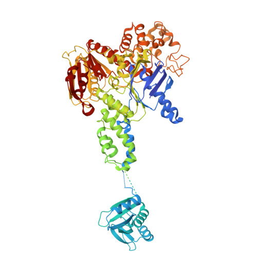
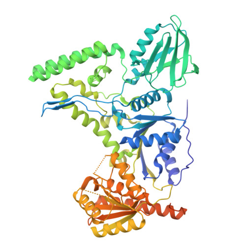
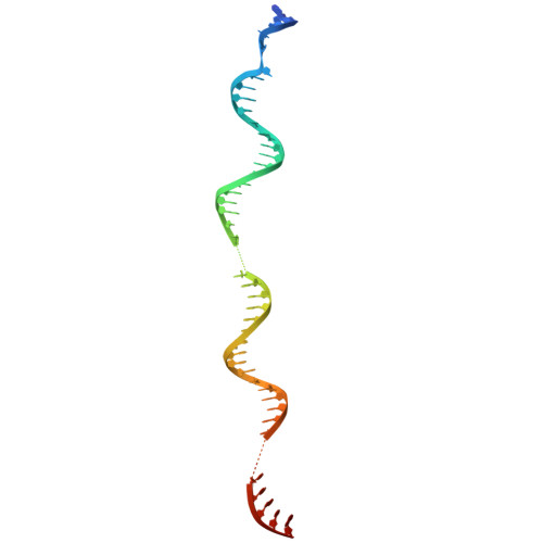
Structural snapshots of the mechanism of ATP-dependent DNA damage recognition by UvrA.

Nirwal, S.Czarnocki-Cieciura, M.Zajko, W.Skowronek, K.Szczepanowski, R.H.Nowotny, M.

(2025) Nat Commun 17: 387-387

  • DOI: https://doi.org/10.1038/s41467-025-67075-y
  • Primary Citation Related Structures: 
    9GXK, 9GXL, 9GXM, 9GXN, 9GXO, 9GXP, 9GXQ, 9GXR, 9GXS, 9GXT, 9GXU, 9GXV, 9GXW, 9GY7

  • PubMed Abstract: 

    Nucleotide excision repair is a DNA repair pathway which detects and fixes various DNA lesions that distort the structure of DNA. In bacteria, the pathway starts with the UvrA protein which has two adenosine triphosphatase modules and forms dimers. The DNA is handed over from UvrA to UvrB, which is a weak helicase that verifies the presence of damage. Despite intense studies, the role of the ATPase activity of UvrA in damage recognition is unclear. Here, we present a series of cryo-electron microscopy structures of UvrA in complex with three different DNAs and in the presence and absence of nucleotides. We also present a structure of UvrA:UvrB:DNA complex. These structures reveal a major rearrangement of the UvrA dimer upon ATP binding. We propose that these conformational changes are used to mechanically probe the integrity of DNA for damage localization. Collectively, our results present snapshots of UvrA's ATP-dependent DNA damage detection.


  • Organizational Affiliation
    • Laboratory of Protein Structure, International Institute of Molecular and Cell Biology, Warsaw, Poland.

Macromolecule Content 

  • Total Structure Weight: 398.72 kDa 
  • Atom Count: 21,176 
  • Modeled Residue Count: 2,726 
  • Deposited Residue Count: 3,346 
  • Unique protein chains: 2
  • Unique nucleic acid chains: 1

Macromolecules


Find similar proteins by:|  3D Structure
Entity ID: 1
MoleculeChains  Sequence LengthOrganismDetailsImage
UvrABC system protein A
A, B
962Acetivibrio thermocellusMutation(s): 0 
Gene Names: uvrACthe_0311
UniProt
Find proteins for A3DC70 (Acetivibrio thermocellus (strain ATCC 27405 / DSM 1237 / JCM 9322 / NBRC 103400 / NCIMB 10682 / NRRL B-4536 / VPI 7372))
Explore A3DC70 
Go to UniProtKB:  A3DC70
Entity Groups
Sequence Clusters30% Identity50% Identity70% Identity90% Identity95% Identity100% Identity
UniProt GroupA3DC70
Sequence Annotations
Expand
Reference Sequence
Find similar proteins by:|  3D Structure
Entity ID: 3
MoleculeChains  Sequence LengthOrganismDetailsImage
UvrABC system protein B
E, F
661Acetivibrio thermocellusMutation(s): 0 
Gene Names: uvrBCthe_0309
UniProt
Find proteins for A3DC68 (Acetivibrio thermocellus (strain ATCC 27405 / DSM 1237 / JCM 9322 / NBRC 103400 / NCIMB 10682 / NRRL B-4536 / VPI 7372))
Explore A3DC68 
Go to UniProtKB:  A3DC68
Entity Groups
Sequence Clusters30% Identity50% Identity70% Identity90% Identity95% Identity100% Identity
UniProt GroupA3DC68
Sequence Annotations
Expand
Reference Sequence
Find similar nucleic acids by:  (by identity cutoff) 
Entity ID: 2
MoleculeChains LengthOrganismImage
DNA (50-MER) with a fluorescein modification - (random sequence built in model)
C, D
50synthetic construct
Sequence Annotations
Expand
Reference Sequence

Small Molecules

Ligands 3 Unique
IDChains Name / Formula / InChI Key2D Diagram3D Interactions
ANP
(Subject of Investigation/LOI)

Query on ANP


Download:Ideal Coordinates CCD File
H [auth A],
I [auth A],
L [auth B],
M [auth B]
PHOSPHOAMINOPHOSPHONIC ACID-ADENYLATE ESTER
C10 H17 N6 O12 P3
PVKSNHVPLWYQGJ-KQYNXXCUSA-N
ZN
(Subject of Investigation/LOI)

Query on ZN


Download:Ideal Coordinates CCD File
G [auth A],
K [auth B]
ZINC ION
Zn
PTFCDOFLOPIGGS-UHFFFAOYSA-N
MG
(Subject of Investigation/LOI)

Query on MG


Download:Ideal Coordinates CCD File
J [auth A],
N [auth B]
MAGNESIUM ION
Mg
JLVVSXFLKOJNIY-UHFFFAOYSA-N

Experimental Data & Validation

Experimental Data

  • Method: ELECTRON MICROSCOPY
  • Resolution: 3.18 Å
  • Aggregation State: PARTICLE 
  • Reconstruction Method: SINGLE PARTICLE 
EM Software:
TaskSoftware PackageVersion
RECONSTRUCTIONcryoSPARC4.5
MODEL REFINEMENTPHENIX1.21.2-5419

Structure Validation

View Full Validation Report


Entry History 

& Funding Information

Deposition Data


Funding OrganizationLocationGrant Number
Polish National Science CentrePoland2017/26/A/NZ1/01098

Revision History  (Full details and data files)

  • Version 1.0: 2025-12-24
    Type: Initial release
  • Version 1.1: 2026-01-21
    Changes: Data collection, Database references