9GTI | pdb_00009gti

X-ray crystal structure of mouse NPTN N-terminal domain


Experimental Data Snapshot

  • Method: X-RAY DIFFRACTION
  • Resolution: 2.03 Å
  • R-Value Free: 
    0.248 (Depositor), 0.259 (DCC) 
  • R-Value Work: 
    0.215 (Depositor), 0.227 (DCC) 
  • R-Value Observed: 
    0.217 (Depositor) 

Starting Model: in silico
View more details

wwPDB Validation   3D Report Full Report


Ligand Structure Quality Assessment 


This is version 1.2 of the entry. See complete history


Literature

Molecular mechanism of ultrafast transport by plasma membrane Ca 2+ -ATPases.

Vinayagam, D.Sitsel, O.Schulte, U.Constantin, C.E.Oosterheert, W.Prumbaum, D.Zolles, G.Fakler, B.Raunser, S.

(2025) Nature 646: 236-245

  • DOI: https://doi.org/10.1038/s41586-025-09402-3
  • Primary Citation of Related Structures:  
    9GSD, 9GSE, 9GSF, 9GSG, 9GSH, 9GSI, 9GSY, 9GTB, 9GTI

  • PubMed Abstract: 

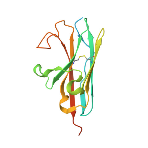
    Tight control of intracellular Ca 2+ levels is fundamental as they are used to control numerous signal transduction pathways 1 . Plasma membrane Ca 2+ -ATPases (PMCAs) have a crucial role in this process by extruding Ca 2+ against a steep concentration gradient from the cytosol to the extracellular space 2 . Although new details of PMCA biology are constantly being uncovered, the structural basis of the most distinguishing features of these pumps, namely, transport rates in the kilohertz range and regulation of activity by the plasma membrane phospholipid PtdIns(4,5)P 2 , has so far remained elusive. Here we present the structures of mouse PMCA2 in the presence and absence of its accessory subunit neuroplastin in eight different stages of its transport cycle. Combined with whole-cell recordings that accurately track PMCA-mediated Ca 2+ extrusion in intact cells, these structures enable us to establish the first comprehensive transport model for a PMCA, reveal the role of disease-causing mutations and uncover the structural underpinnings of regulatory PMCA-phospholipid interaction. The transport cycle-dependent dynamics of PtdIns(4,5)P 2 are fundamental for its role as a 'latch' promoting the fast release of Ca 2+ and opening a passageway for counter-ions. These actions are required for maintaining the ultra-fast transport cycle. Moreover, we identify the PtdIns(4,5)P 2 -binding site as an unanticipated target for drug-mediated manipulation of intracellular Ca 2+ levels. Our work provides detailed structural insights into the uniquely fast operation of native PMCA-type Ca 2+ pumps and its control by membrane lipids and drugs.


  • Organizational Affiliation
    • Department of Structural Biochemistry, Max Planck Institute of Molecular Physiology, Dortmund, Germany.

Macromolecules
Find similar proteins by:  (by identity cutoff)  |  3D Structure
Entity ID: 1
MoleculeChains Sequence LengthOrganismDetailsImage
Neuroplastin
A, B, C, D
161Mus musculusMutation(s): 0 
Gene Names: NptnSdfr1Sdr1
UniProt & NIH Common Fund Data Resources
Find proteins for P97300 (Mus musculus)
Explore P97300 
Go to UniProtKB:  P97300
IMPC:  MGI:108077
Entity Groups  
Sequence Clusters30% Identity50% Identity70% Identity90% Identity95% Identity100% Identity
UniProt GroupP97300
Sequence Annotations
Expand
  • Reference Sequence
Small Molecules
Ligands 2 Unique
IDChains Name / Formula / InChI Key2D Diagram3D Interactions
EPE (Subject of Investigation/LOI)
Query on EPE

Download Ideal Coordinates CCD File 
T [auth D]4-(2-HYDROXYETHYL)-1-PIPERAZINE ETHANESULFONIC ACID
C8 H18 N2 O4 S
JKMHFZQWWAIEOD-UHFFFAOYSA-N
PEG (Subject of Investigation/LOI)
Query on PEG

Download Ideal Coordinates CCD File 
E [auth A]
F [auth A]
G [auth A]
H [auth A]
I [auth A]
E [auth A],
F [auth A],
G [auth A],
H [auth A],
I [auth A],
J [auth B],
K [auth B],
L [auth B],
M [auth B],
N [auth C],
O [auth C],
P [auth C],
Q [auth C],
R [auth D],
S [auth D]
DI(HYDROXYETHYL)ETHER
C4 H10 O3
MTHSVFCYNBDYFN-UHFFFAOYSA-N
Experimental Data & Validation

Experimental Data

  • Method: X-RAY DIFFRACTION
  • Resolution: 2.03 Å
  • R-Value Free:  0.248 (Depositor), 0.259 (DCC) 
  • R-Value Work:  0.215 (Depositor), 0.227 (DCC) 
  • R-Value Observed: 0.217 (Depositor) 
Space Group: P 32 2 1
Unit Cell:
Length ( Å )Angle ( ˚ )
a = 85.907α = 90
b = 85.907β = 90
c = 163.285γ = 120
Software Package:
Software NamePurpose
PHENIXrefinement
XDSdata reduction
XSCALEdata scaling
PHASERphasing

Structure Validation

View Full Validation Report


Ligand Structure Quality Assessment 


Entry History & Funding Information

Deposition Data


Funding OrganizationLocationGrant Number
Max Planck SocietyGermany--
German Research Foundation (DFG)GermanyTRR 152/3, number 239283807 project P02

Revision History  (Full details and data files)

  • Version 1.0: 2025-08-06
    Type: Initial release
  • Version 1.1: 2025-09-03
    Changes: Database references
  • Version 1.2: 2025-10-15
    Changes: Database references