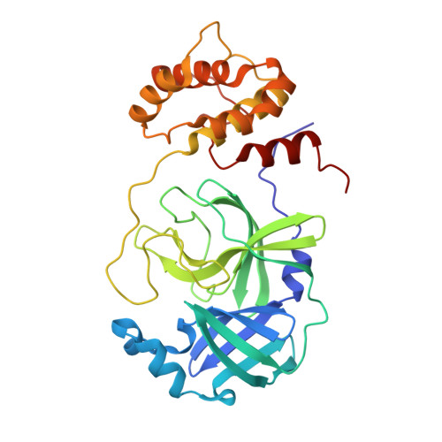
9GHN | pdb_00009ghn

Structure of SARS-CoV-2 Main Protease (Mpro) with mutation of Q256A


Experimental Data Snapshot

  • Method: X-RAY DIFFRACTION
  • Resolution: 1.40 Å
  • R-Value Free: 
    0.218 (Depositor), 0.195 (DCC) 
  • R-Value Work: 
    0.193 (Depositor) 
  • R-Value Observed: 
    0.193 (Depositor) 

Starting Model: experimental
View more details

wwPDB Validation 3D Report Full Report

Validation slider image for 9GHN

This is version 1.1 of the entry. See complete history. 

Literature

Statistical crystallography reveals an allosteric network in SARS-CoV-2 M pro.

Creon, A., Scheer, T.E.S., Reinke, P., Rahmani Mashhour, A., Gunther, S., Niebling, S., Schamoni-Kast, K., Uetrecht, C., Meents, A., Chapman, H.N., Sprenger, J., Lane, T.J.

(2026) Commun Biol 9

  • DOI: https://doi.org/10.1038/s42003-026-10127-w
  • Primary Citation Related Structures: 
    9GHN, 9GHO, 9GI6

  • PubMed Abstract: 

    To interpret and transmit biological signals, proteins use correlated motions. Experimental determination of these dynamics and the structural distributions they generate remains a key challenge. Here, using 1146 crystal structures of the main protease (M pro ) from SARS-CoV-2, we were able to infer a model of the enzyme's structural fluctuations. M pro is regulated by concentration, becoming enzymatically active after forming a homodimer. To understand the structural changes that enable dimerization to activate catalysis, we employed our model, predicting which regions of the dimerization domain are structurally correlated with the active site. Mutations at these positions, expected to disrupt catalysis, resulted in a dramatic reduction in activity in one case, a mild effect in the second, and none in the third. Additional crystallography and biophysical experiments provide a mechanistic explanation for these results. Our work suggests that a statistical crystallography, in which numerous crystallographic datasets are analyzed, can reveal the structural fluctuations of protein native states and help uncover their biological function.


  • Organizational Affiliation: 
    • Center for Free-Electron Laser Science CFEL, Deutsches Elektronen-Synchrotron DESY, Hamburg, Germany.

Macromolecule Content 

  • Total Structure Weight: 67.84 kDa 
  • Atom Count: 5,537 
  • Modeled Residue Count: 612 
  • Deposited Residue Count: 612 
  • Unique protein chains: 1

Macromolecules

Find similar proteins by:|  3D Structure
Entity ID: 1
MoleculeChains  Sequence LengthOrganismDetailsImage
3C-like proteinase nsp5
A, B
306Severe acute respiratory syndrome coronavirus 2Mutation(s): 1 
EC: 3.4.22.69
UniProt
Find proteins for P0DTC1 (Severe acute respiratory syndrome coronavirus 2)
Explore P0DTC1 
Go to UniProtKB:  P0DTC1
Entity Groups
Sequence Clusters30% Identity50% Identity70% Identity90% Identity95% Identity100% Identity
UniProt GroupP0DTC1
Sequence Annotations
Expand
Reference Sequence

Small Molecules

Ligands 3 Unique
IDChains Name / Formula / InChI Key2D Diagram3D Interactions
EDO

Query on EDO


Download:Ideal Coordinates CCD File
C [auth A],
F [auth B],
G [auth B]
1,2-ETHANEDIOL
C2 H6 O2
LYCAIKOWRPUZTN-UHFFFAOYSA-N
CL

Query on CL


Download:Ideal Coordinates CCD File
D [auth A],
H [auth B]
CHLORIDE ION
Cl
VEXZGXHMUGYJMC-UHFFFAOYSA-M
NA

Query on NA


Download:Ideal Coordinates CCD File
E [auth A],
I [auth B]
SODIUM ION
Na
FKNQFGJONOIPTF-UHFFFAOYSA-N

Experimental Data & Validation

Experimental Data

  • Method: X-RAY DIFFRACTION
  • Resolution: 1.40 Å
  • R-Value Free:  0.218 (Depositor), 0.195 (DCC) 
  • R-Value Work:  0.193 (Depositor) 
  • R-Value Observed: 0.193 (Depositor) 
Space Group: P 21 21 21
Unit Cell:
Length ( Å )Angle ( ˚ )
a = 78.538α = 90
b = 88.438β = 90
c = 100.766γ = 90
Software Package:
Software NamePurpose
PHENIXrefinement
PHENIXrefinement
DIALSdata reduction
DIALSdata scaling
PHASERphasing

Structure Validation

View Full Validation Report


Entry History 

& Funding Information

Deposition Data


Funding OrganizationLocationGrant Number
Helmholtz AssociationGermany--

Revision History  (Full details and data files)

  • Version 1.0: 2025-09-03
    Type: Initial release
  • Version 1.1: 2026-09-23
    Changes: Database references