Structure of the T=13 capsid of infectious pancreatic necrosis virus (IPNV)-a salmonid birnavirus.
Munke, A., Ahmed Abdelrahim Gamil, A., Mikalsen, A.B., Wang, H., Evensen, O., Okamoto, K.(2025) J Virol 99: e0145424-e0145424
- PubMed: 39817769 Search on PubMedSearch on PubMed Central
- DOI: https://doi.org/10.1128/jvi.01454-24
- Primary Citation Related Structures:
9GG2 - PubMed Abstract:
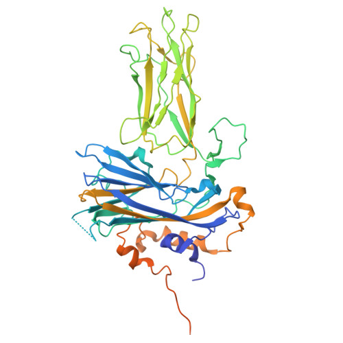
Birnaviruses infect a broad range of vertebrate hosts, including fish and birds, and cause substantial economic losses in the fishery and livestock industries. The infectious pancreatic necrosis virus (IPNV), an aquabirnavirus, specifically infects salmonids. While structures on T=1 subviral particles of the birnaviruses, including IPNV, have been studied, structural insights into the infectious T=13 particles have been limited to the infectious bursal disease virus (IBDV), an avibirnavirus. Determining the capsid structure of the T=13 particle of IPNV is crucial for advancing knowledge of its antigenicity, capsid assembly, and possible functional structures. Here, the capsid structure of the IPNV L5 strain has been determined at a resolution of 2.75 Å. The overall structure resembles the T=13 IBDV structure, with notable differences in the surface loops on the P domain of the VP2 capsid protein essential for antigenicity and virulence. Additionally, previously undescribed structural features have been identified, including the C-terminal regions of the VP2 subunits within the pentagonal assembly unit at each 5-fold axis, which interlock with adjacent VP2 subunits. This interlocking, together with class-averaged projections of triangular and pentagonal units, suggests that the pentagonal unit formation could be important for a correct T=13 particle assembly, preventing the formation of T=1 subviral particles. Furthermore, positively charged residues in obstructed capsid pores at each 5-fold axis are speculated to facilitate intraparticle genome synthesis of IPNV.IMPORTANCEAquabirnaviruses cause deadly infectious diseases in salmonid fish, posing significant challenges for both wild and farmed fish populations. The most prevalent aquabirnavirus worldwide is the infectious pancreatic necrosis virus, whose multifunctional capsid is critical to its infection, replication, and maturation. Previously, research has focused on the structure of the virus' non-infectious subviral capsid. In this study, however, the first structure of the large, infectious, and functional form of the capsid has been determined. This new capsid structure reveals functional motifs that were previously unclear in the non-infectious capsid. These motifs are believed to be essential for the virus' replication and particle assembly, making them promising targets for developing strategies to control virus proliferation.
- Laboratory of Molecular Biophysics, Department of Cell and Molecular Biology, Uppsala University, Uppsala, Sweden.
Organizational Affiliation: