


Crystal structure of HEI10
Milburn, A.E., Espejo-Serrano, C., Dunce, J.M., Davies, O.R.To be published.
Experimental Data Snapshot
Starting Model: in silico
View more details
wwPDB Validation 3D Report Full Report

Entity ID: 1 | |||||
|---|---|---|---|---|---|
| Molecule | Chains | Sequence Length | Organism | Details | Image |
| E3 ubiquitin-protein ligase CCNB1IP1 | 198 | Homo sapiens | Mutation(s): 0 Gene Names: CCNB1IP1, C14orf18, HEI10 EC: 2.3.2.27 | 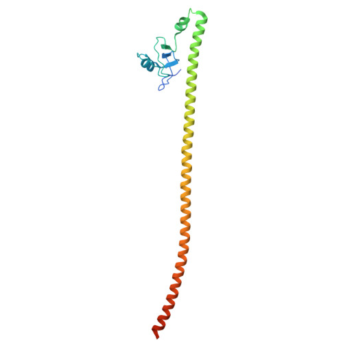 | |
UniProt & NIH Common Fund Data Resources | |||||
PHAROS: Q9NPC3 GTEx: ENSG00000100814 | |||||
Entity Groups | |||||
| Sequence Clusters | 30% Identity50% Identity70% Identity90% Identity95% Identity100% Identity | ||||
| UniProt Group | Q9NPC3 | ||||
Sequence AnnotationsExpand | |||||
Reference Sequence | |||||
| Ligands 1 Unique | |||||
|---|---|---|---|---|---|
| ID | Chains | Name / Formula / InChI Key | 2D Diagram | 3D Interactions | |
| ZN (Subject of Investigation/LOI) Download:Ideal Coordinates CCD File | E [auth A] F [auth A] G [auth B] H [auth B] I [auth C] | ZINC ION Zn PTFCDOFLOPIGGS-UHFFFAOYSA-N |  | ||
| Length ( Å ) | Angle ( ˚ ) |
|---|---|
| a = 53.198 | α = 90 |
| b = 73.857 | β = 99.276 |
| c = 124.032 | γ = 90 |
| Software Name | Purpose |
|---|---|
| PHENIX | refinement |
| autoPROC | data reduction |
| STARANISO | data scaling |
| PHASER | phasing |
| Funding Organization | Location | Grant Number |
|---|---|---|
| Wellcome Trust | United Kingdom | 219413/Z/19/Z |


