Fragment Discovery by X-Ray Crystallographic Screening Targeting the CTP Binding Site of Pseudomonas Aeruginosa IspD.
Willocx, D., D'Auria, L., Walsh, D., Scherer, H., Alhayek, A., Hamed, M.M., Borel, F., Diamanti, E., Hirsch, A.K.H.(2025) Angew Chem Int Ed Engl 64: e202414615-e202414615
- PubMed: 39676054 Search on PubMedSearch on PubMed Central
- DOI: https://doi.org/10.1002/anie.202414615
- Primary Citation Related Structures:
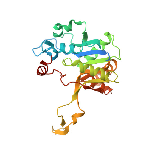
9GBY, 9GC8, 9GCA - PubMed Abstract:
With antimicrobial resistance (AMR) reaching alarming levels, new anti-infectives with unprecedented mechanisms of action are urgently needed. The 2-C-methylerythritol-D-erythritol-4-phosphate (MEP) pathway represents an attractive source of drug targets due to its essential role in numerous pathogenic Gram-negative bacteria and Mycobacterium tuberculosis (Mt), whilst being absent in human cells. Here, we solved the first crystal structure of Pseudomonas aeruginosa (Pa) IspD, the third enzyme in the MEP pathway and present the discovery of a fragment-based compound class identified through crystallographic screening of PaIspD. The initial fragment occupies the CTP binding cavity within the active site. Confirmation of fragment-protein interactions was achieved through 1 H saturation-transfer difference nuclear magnetic resonance ( 1 H-STD NMR spectroscopy). Building upon these findings and insights from the co-crystal structures, we identified two growth vectors for fragment growing. We synthesized derivatives addressing both growth vectors, which showed improved affinities for PaIspD. Our new fragment class targets PaIspD, displays promising affinity and favorable growth vectors for further optimization.
- Helmholtz Institute for Pharmaceutical Research Saarland (HIPS), Helmholtz Centre for Infection Research (HZI), Campus E8.1, 66123, Saarbrücken, Germany.
Organizational Affiliation: