9G6D | pdb_00009g6d

CLC7/OSTM1 complex in the absence of PIP2 lipid.


Experimental Data Snapshot

  • Method: ELECTRON MICROSCOPY
  • Resolution: 2.70 Å
  • Aggregation State: PARTICLE 
  • Reconstruction Method: SINGLE PARTICLE 

wwPDB Validation 3D Report Full Report

Validation slider image for 9G6D

This is version 1.1 of the entry. See complete history

Literature

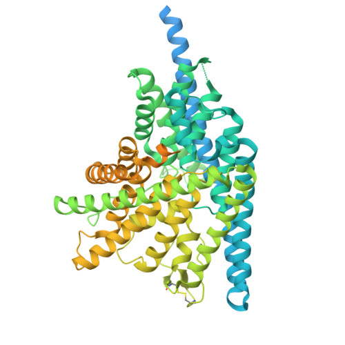
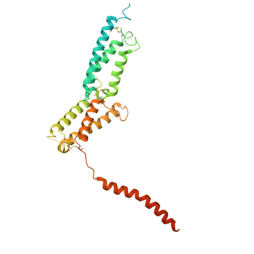
Mechanism of phosphoinositide regulation of lysosomal pH via inhibition of CLC-7.

Hilton, J.K.Lin, Y.Sefah, E.Deme, J.C.Parker, J.L.Langton, M.J.Grabe, M.Lea, S.Newstead, S.Mindell, J.A.

(2025) bioRxiv 

  • DOI: https://doi.org/10.1101/2025.10.01.679551
  • Primary Citation Related Structures: 
    9G6C, 9G6D, 9G6E

  • PubMed Abstract: 

    Lysosomes process cellular waste and coordinate responses to metabolic challenge. Central to lysosomal homeostasis are phosphoinositide lipids, key signaling molecules which establish organelle identity, regulate membrane dynamics and are tightly linked to the pathophysiology and therapy of lysosomal storage disorders, neurodegeneration, and cancer. Phosphatidylinositol 3,5-bisphosphate (PI(3,5)P2) interacts with multiple lysosomal membrane proteins and plays a critical role in regulating lysosomal pH by directly inhibiting the chloride/proton antiporter ClC-7, though the molecular mechanism of this inhibition remains unclear. Here, using a combination of functional, structural, and computational analysis, we demonstrate that PI(3,5)P2 binding dramatically remodels the structure of ClC-7 by inducing close association between cytosolic and transmembrane domains. Disease-causing mutations show increased transport activity through loss of PI(3,5)P2 binding and subsequent inhibition. Conversely, ClC-7 activation is correlated with dissociation and increased disorder of the cytoplasmic domain along with novel transmembrane domain conformations, revealing a mechanistic link between specific lysosomal lipids, transporter regulation, and the enigmatic basis of the ClC-7 slow gate.


  • Organizational Affiliation
    • Membrane Transport Biophysics Section, National Institute of Neurological Disorders and Stroke, NIH, Bethesda, MD 20892, USA.

Macromolecule Content 

  • Total Structure Weight: 257.08 kDa 
  • Atom Count: 11,292 
  • Modeled Residue Count: 1,380 
  • Deposited Residue Count: 2,278 
  • Unique protein chains: 2

Macromolecules

Find similar proteins by:|  3D Structure
Entity ID: 1
MoleculeChains  Sequence LengthOrganismDetailsImage
H(+)/Cl(-) exchange transporter 7
A, C
805Homo sapiensMutation(s): 0 
Gene Names: CLCN7
Membrane Entity: Yes 
UniProt & NIH Common Fund Data Resources
Find proteins for P51798 (Homo sapiens)
Explore P51798 
Go to UniProtKB:  P51798
PHAROS:  P51798
GTEx:  ENSG00000103249 
Entity Groups
Sequence Clusters30% Identity50% Identity70% Identity90% Identity95% Identity100% Identity
UniProt GroupP51798
Sequence Annotations
Expand
Reference Sequence
Find similar proteins by:|  3D Structure
Entity ID: 2
MoleculeChains  Sequence LengthOrganismDetailsImage
Osteopetrosis-associated transmembrane protein 1
B, D
334Homo sapiensMutation(s): 0 
Gene Names: OSTM1GLHSPC019UNQ6098/PRO21201
Membrane Entity: Yes 
UniProt & NIH Common Fund Data Resources
Find proteins for Q86WC4 (Homo sapiens)
Explore Q86WC4 
Go to UniProtKB:  Q86WC4
PHAROS:  Q86WC4
GTEx:  ENSG00000081087 
Entity Groups
Sequence Clusters30% Identity50% Identity70% Identity90% Identity95% Identity100% Identity
UniProt GroupQ86WC4
Sequence Annotations
Expand
Reference Sequence

Oligosaccharides

Help  
Entity ID: 3
MoleculeChains Length2D Diagram GlycosylationD Interactions
alpha-D-mannopyranose-(1-3)-[alpha-D-mannopyranose-(1-6)]beta-D-mannopyranose-(1-4)-2-acetamido-2-deoxy-beta-D-glucopyranose-(1-4)-2-acetamido-2-deoxy-beta-D-glucopyranoseE,
F [auth H]
5N/A
Glycosylation Resources
GlyTouCan: G22768VO
GlyCosmos: G22768VO
GlyGen: G22768VO

Small Molecules

Ligands 2 Unique
IDChains Name / Formula / InChI Key2D Diagram3D Interactions
Y01

Query on Y01


Download:Ideal Coordinates CCD File
J [auth A]
K [auth A]
L [auth A]
P [auth C]
Q [auth C]
J [auth A],
K [auth A],
L [auth A],
P [auth C],
Q [auth C],
R [auth C]
CHOLESTEROL HEMISUCCINATE
C31 H50 O4
WLNARFZDISHUGS-MIXBDBMTSA-N
CL
(Subject of Investigation/LOI)

Query on CL


Download:Ideal Coordinates CCD File
G [auth A]
H [auth A]
I [auth A]
M [auth C]
N [auth C]
G [auth A],
H [auth A],
I [auth A],
M [auth C],
N [auth C],
O [auth C]
CHLORIDE ION
Cl
VEXZGXHMUGYJMC-UHFFFAOYSA-M

Experimental Data & Validation

Experimental Data

  • Method: ELECTRON MICROSCOPY
  • Resolution: 2.70 Å
  • Aggregation State: PARTICLE 
  • Reconstruction Method: SINGLE PARTICLE 

Structure Validation

View Full Validation Report


Entry History 

& Funding Information

Deposition Data


Funding OrganizationLocationGrant Number
Wellcome TrustUnited Kingdom--

Revision History  (Full details and data files)

  • Version 1.0: 2025-07-30
    Type: Initial release
  • Version 1.1: 2025-12-10
    Changes: Data collection, Database references