9G4T | pdb_00009g4t

Beta carbonic anhydrase CsoSCA from the Halothiobacillus neapolitanus alpha-carboxysome


Experimental Data Snapshot

  • Method: ELECTRON MICROSCOPY
  • Resolution: 2.51 Å
  • Aggregation State: PARTICLE 
  • Reconstruction Method: SINGLE PARTICLE 

Starting Model: in silico
View more details

wwPDB Validation   3D Report Full Report


This is version 1.1 of the entry. See complete history


Literature

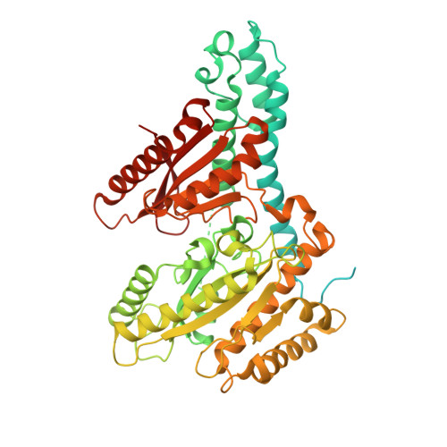
Structure and encapsulation of carbonic anhydrase within the alpha-carboxysome.

Ng, P.C.Adegbite, O.Li, T.Basle, A.Marles-Wright, J.Liu, L.N.

(2025) Proc Natl Acad Sci U S A 122: e2523723122-e2523723122

  • DOI: https://doi.org/10.1073/pnas.2523723122
  • Primary Citation of Related Structures:  
    9G4T, 9GVC, 9GW1

  • PubMed Abstract: 

    Carboxysomes in cyanobacteria and certain proteobacteria enable efficient CO 2 fixation by encapsulating ribulose-1,5-bisphosphate carboxylase/oxygenase (Rubisco) and carbonic anhydrase (CA) within a semipermeable shell. Sequestered CA catalyze the rapid interconversion of CO 2 and HCO 3 - , supplying elevated levels of CO 2 to boost Rubisco carboxylation. Despite its essential role, the structure and encapsulation of CA within carboxysomes remain poorly understood. Here, we determined the molecular structure of α-carboxysomal CA from the model chemoautotrophic bacterium Halothiobacillus neapolitanus ( Hn CsoSCA). Hn CsoSCA adopts a trimer-of-dimers oligomeric structure without the incorporation of a zinc ion at its symmetric center. Using synthetic minishells, we demonstrate that Hn CsoSCA interacts with the CsoS1A shell hexamer and is incorporated into the minishells at the inner surface, independent of the CsoS2 linker protein. Hn CsoSCA truncations suggest nonspecific interactions between Hn CsoSCA and CsoS1A. We further show that Hn CsoSCA bridges Rubisco and the shell facets. Our study offers insights into the assembly and encapsulation mechanisms of α-carboxysomes and provides the framework for reprogramming carboxysome structures for synthetic biology and biotechnological applications.


  • Organizational Affiliation
    • Institute of Systems, Molecular and Integrative Biology, University of Liverpool, Liverpool L69 7ZB, United Kingdom.

Macromolecules
Find similar proteins by:  (by identity cutoff)  |  3D Structure
Entity ID: 1
MoleculeChains Sequence LengthOrganismDetailsImage
Immunoglobulin G-binding protein G,Carboxysome shell carbonic anhydrase614Streptococcus sp. 'group GHalothiobacillus neapolitanus
This entity is chimeric
Mutation(s): 0 
Gene Names: spgcsoS3ORF1Hneap_0919
EC: 4.2.1.1
UniProt
Find proteins for O85042 (Halothiobacillus neapolitanus (strain ATCC 23641 / c2))
Explore O85042 
Go to UniProtKB:  O85042
Find proteins for P06654 (Streptococcus sp. group G)
Explore P06654 
Go to UniProtKB:  P06654
Entity Groups  
Sequence Clusters30% Identity50% Identity70% Identity90% Identity95% Identity100% Identity
UniProt GroupsO85042P06654
Sequence Annotations
Expand
  • Reference Sequence
Small Molecules
Ligands 1 Unique
IDChains Name / Formula / InChI Key2D Diagram3D Interactions
ZN
Query on ZN

Download Ideal Coordinates CCD File 
B [auth A]ZINC ION
Zn
PTFCDOFLOPIGGS-UHFFFAOYSA-N
Experimental Data & Validation

Experimental Data

  • Method: ELECTRON MICROSCOPY
  • Resolution: 2.51 Å
  • Aggregation State: PARTICLE 
  • Reconstruction Method: SINGLE PARTICLE 
EM Software:
TaskSoftware PackageVersion
RECONSTRUCTIONcryoSPARC4.5
MODEL REFINEMENTPHENIX1.21.1

Structure Validation

View Full Validation Report


Entry History & Funding Information

Deposition Data


Funding OrganizationLocationGrant Number
Biotechnology and Biological Sciences Research Council (BBSRC)United Kingdom2599456

Revision History  (Full details and data files)

  • Version 1.0: 2025-07-30
    Type: Initial release
  • Version 1.1: 2025-11-26
    Changes: Data collection, Database references