9FND | pdb_00009fnd

Transcriptional activator PafBC bound to mycobacterial RNA polymerase


Experimental Data Snapshot

  • Method: ELECTRON MICROSCOPY
  • Resolution: 4.00 Å
  • Aggregation State: PARTICLE 
  • Reconstruction Method: SINGLE PARTICLE 

Starting Model: in silico
View more details

wwPDB Validation   3D Report Full Report


This is version 1.1 of the entry. See complete history


Literature

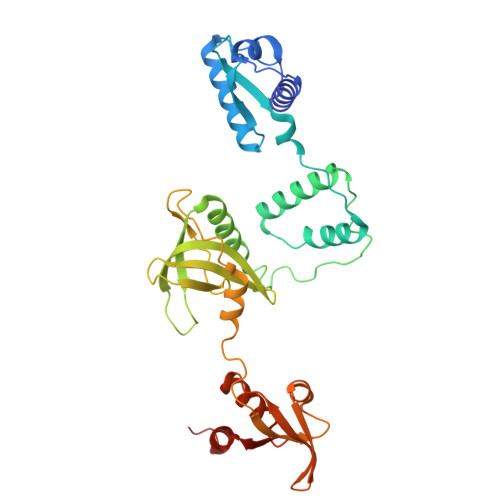
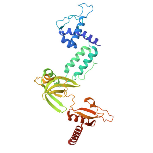
Single-stranded DNA binding to the transcription factor PafBC triggers the mycobacterial DNA damage response.

Schilling, C.M.Zdanowicz, R.Rabl, J.Muller, A.U.Boehringer, D.Glockshuber, R.Weber-Ban, E.

(2025) Sci Adv 11: eadq9054-eadq9054

  • DOI: https://doi.org/10.1126/sciadv.adq9054
  • Primary Citation of Related Structures:  
    9FND, 9FNE

  • PubMed Abstract: 

    The DNA damage response in mycobacteria is controlled by the heterodimeric transcription factor PafBC, a member of the WYL domain-containing protein family. It has been shown that PafBC induces transcription of its regulon by reprogramming the housekeeping RNA polymerase holoenzyme to recognize PafBC-dependent promoters through sigma adaptation. However, the mechanism by which DNA damage is sensed and translated into PafBC activation has remained unclear. Here, we demonstrate that the binding of single-stranded DNA (ssDNA) to the WYL domains of PafBC activates the transcription factor. Our cryo-electron microscopy structure of full-length PafBC in its active conformation, bound to the transcription initiation complex, reveals a previously unknown mode of interaction between the ssDNA and the WYL domains. Using biochemical experiments, we show that short ssDNA fragments bind to PafBC dynamically, resulting in deactivation as ssDNA levels decrease postrepair. Our findings shed light on the mechanism linking DNA damage to PafBC activation and expand our understanding of WYL domain-containing proteins.


  • Organizational Affiliation
    • ETH Zurich, Institute of Molecular Biology and Biophysics, 8093 Zurich, Switzerland.

Macromolecules
Find similar proteins by:  (by identity cutoff)  |  3D Structure
Entity ID: 1
MoleculeChains Sequence LengthOrganismDetailsImage
Transcriptional regulator-like proteinA [auth X]333Mycolicibacterium smegmatis MC2 155Mutation(s): 0 
Gene Names: pafBMSMEI_3799
UniProt
Find proteins for I7G3U5 (Mycolicibacterium smegmatis (strain ATCC 700084 / mc(2)155))
Explore I7G3U5 
Go to UniProtKB:  I7G3U5
Entity Groups  
Sequence Clusters30% Identity50% Identity70% Identity90% Identity95% Identity100% Identity
UniProt GroupI7G3U5
Sequence Annotations
Expand
  • Reference Sequence
Find similar proteins by:  (by identity cutoff)  |  3D Structure
Entity ID: 2
MoleculeChains Sequence LengthOrganismDetailsImage
PafCB [auth Y]318Mycolicibacterium smegmatis MC2 155Mutation(s): 0 
Gene Names: MSMEG_3888
UniProt
Find proteins for A0QZ41 (Mycolicibacterium smegmatis (strain ATCC 700084 / mc(2)155))
Explore A0QZ41 
Go to UniProtKB:  A0QZ41
Entity Groups  
Sequence Clusters30% Identity50% Identity70% Identity90% Identity95% Identity100% Identity
UniProt GroupA0QZ41
Sequence Annotations
Expand
  • Reference Sequence
Experimental Data & Validation

Experimental Data

  • Method: ELECTRON MICROSCOPY
  • Resolution: 4.00 Å
  • Aggregation State: PARTICLE 
  • Reconstruction Method: SINGLE PARTICLE 
EM Software:
TaskSoftware PackageVersion
RECONSTRUCTIONcryoSPARC

Structure Validation

View Full Validation Report


Entry History & Funding Information

Deposition Data


Funding OrganizationLocationGrant Number
Swiss National Science FoundationSwitzerland310030_215606

Revision History  (Full details and data files)

  • Version 1.0: 2025-02-12
    Type: Initial release
  • Version 1.1: 2025-02-19
    Changes: Data collection, Database references