9FL8 | pdb_00009fl8

Stapled peptide bound to NOT9-NOT1 complex


Experimental Data Snapshot

  • Method: X-RAY DIFFRACTION
  • Resolution: 2.64 Å
  • R-Value Free: 
    0.249 (Depositor), 0.248 (DCC) 
  • R-Value Work: 
    0.203 (Depositor), 0.203 (DCC) 
  • R-Value Observed: 
    0.205 (Depositor) 

Starting Model: experimental
View more details

wwPDB Validation 3D Report Full Report

Validation slider image for 9FL8

This is version 1.0 of the entry. See complete history

Literature

Stapled Peptides as Inhibitors of mRNA Deadenylation.

Pal, S.Gordijenko, I.Schmeing, S.Biswas, S.Akbulut, Y.Gasper, R.'t Hart, P.

(2025) Angew Chem Int Ed Engl 64: e202413911-e202413911

  • DOI: https://doi.org/10.1002/anie.202413911
  • Primary Citation Related Structures: 
    9FL8

  • PubMed Abstract: 

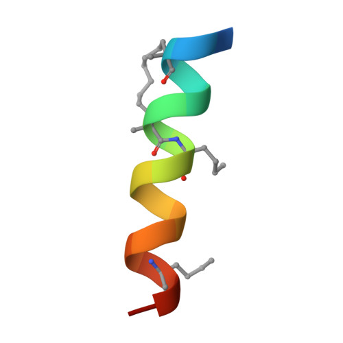
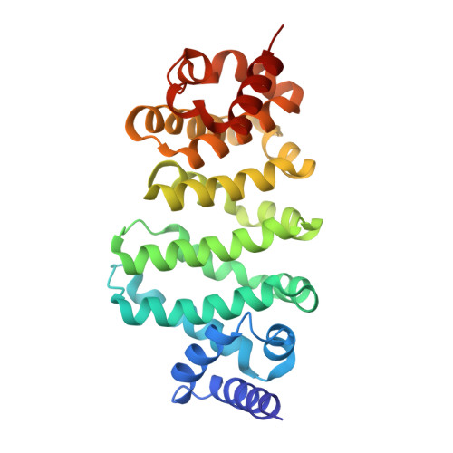
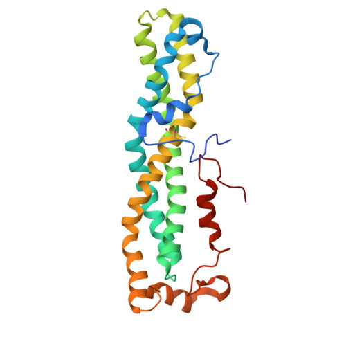
    Therapeutic intervention targeting mRNA typically aims at reducing the levels of disease-causing sequences. Achieving the opposite effect of blocking the destruction of beneficial mRNA remains underexplored. The degradation of mRNA starts with the removal of poly(A) tails, reducing their stability and translational activity, which is mainly regulated by the CCR4-NOT complex. The subunit NOT9 binds various RNA binding proteins, that recruit mRNA in a sequence-specific manner to the CCR4-NOT complex to promote their deadenylation. These RNA binding proteins interact with NOT9 through a helical NOT9 binding motif, which we used as a starting point for development of the hydrocarbon stapled peptide NIP-2. The peptide (K D =60.4 nM) was able to inhibit RNA-binding (IC 50 =333 nM) as well as the deadenylation activity of the CCR4-NOT complex in vitro while being cell-permeable (cell-permeability EC 50 =2.44 μM). A co-crystal structure of NIP-2 bound to NOT9 allowed further optimization of the peptide through point mutation leading to NIP-2-H27A-N 3 (K D =122 nM) with high cell permeability (cell-permeability EC 50 =0.34 μM). The optimized peptide was able to inhibit deadenylation of target mRNAs when used in HeLa cells at a concentration of 100 μM, demonstrating the feasibility of increasing mRNA stability.


  • Organizational Affiliation
    • Chemical Genomics Centre, Max Planck Institute of Molecular Physiology, Otto-Hahn-Strasse 11, 44227, Dortmund, Germany.

Macromolecule Content 

  • Total Structure Weight: 122.05 kDa 
  • Atom Count: 8,498 
  • Modeled Residue Count: 1,049 
  • Deposited Residue Count: 1,072 
  • Unique protein chains: 3

Macromolecules

Find similar proteins by:  Sequence   |   3D Structure  
Entity ID: 1
MoleculeChains  Sequence LengthOrganismDetailsImage
Protein bag of marblesA [auth G],
B [auth F]
19Drosophila melanogasterMutation(s): 6 
UniProt
Find proteins for P22745 (Drosophila melanogaster)
Explore P22745 
Go to UniProtKB:  P22745
Entity Groups
Sequence Clusters30% Identity50% Identity70% Identity90% Identity95% Identity100% Identity
UniProt GroupP22745
Sequence Annotations
Expand
Reference Sequence
Find similar proteins by:|  3D Structure
Entity ID: 2
MoleculeChains  Sequence LengthOrganismDetailsImage
CCR4-NOT transcription complex subunit 9C [auth B],
E [auth C]
273Homo sapiensMutation(s): 0 
Gene Names: CNOT9RCD1RQCD1
UniProt & NIH Common Fund Data Resources
Find proteins for Q92600 (Homo sapiens)
Explore Q92600 
Go to UniProtKB:  Q92600
PHAROS:  Q92600
GTEx:  ENSG00000144580 
Entity Groups
Sequence Clusters30% Identity50% Identity70% Identity90% Identity95% Identity100% Identity
UniProt GroupQ92600
Sequence Annotations
Expand
Reference Sequence
Find similar proteins by:|  3D Structure
Entity ID: 3
MoleculeChains  Sequence LengthOrganismDetailsImage
CCR4-NOT transcription complex subunit 1D [auth A],
F [auth D]
244Homo sapiensMutation(s): 0 
Gene Names: CNOT1CDC39KIAA1007NOT1AD-005
UniProt & NIH Common Fund Data Resources
Find proteins for A5YKK6 (Homo sapiens)
Explore A5YKK6 
Go to UniProtKB:  A5YKK6
PHAROS:  A5YKK6
GTEx:  ENSG00000125107 
Entity Groups
Sequence Clusters30% Identity50% Identity70% Identity90% Identity95% Identity100% Identity
UniProt GroupA5YKK6
Sequence Annotations
Expand
Reference Sequence

Experimental Data & Validation

Experimental Data

  • Method: X-RAY DIFFRACTION
  • Resolution: 2.64 Å
  • R-Value Free:  0.249 (Depositor), 0.248 (DCC) 
  • R-Value Work:  0.203 (Depositor), 0.203 (DCC) 
  • R-Value Observed: 0.205 (Depositor) 
Space Group: P 32 2 1
Unit Cell:
Length ( Å )Angle ( ˚ )
a = 106.562α = 90
b = 106.562β = 90
c = 262.572γ = 120
Software Package:
Software NamePurpose
PHENIXrefinement
Aimlessdata scaling
XDSdata reduction
PHASERphasing

Structure Validation

View Full Validation Report


Entry History 

& Funding Information

Deposition Data


Funding OrganizationLocationGrant Number
H2020 Marie Curie Actions of the European CommissionEuropean UnionHORIZON-MSCA-2021-PF-01-101061526

Revision History  (Full details and data files)

  • Version 1.0: 2025-01-29
    Type: Initial release