9F6G | pdb_00009f6g

Human USP30 chimera bound to Ubiquitin-PA


Experimental Data Snapshot

  • Method: X-RAY DIFFRACTION
  • Resolution: 1.50 Å
  • R-Value Free: 
    0.211 (Depositor), 0.211 (DCC) 
  • R-Value Work: 
    0.183 (Depositor), 0.183 (DCC) 
  • R-Value Observed: 
    0.184 (Depositor) 

Starting Models: in silico, experimental
View more details

wwPDB Validation 3D Report Full Report

Validation slider image for 9F6G

Ligand Structure Quality Assessment 


This is version 1.2 of the entry. See complete history

Literature

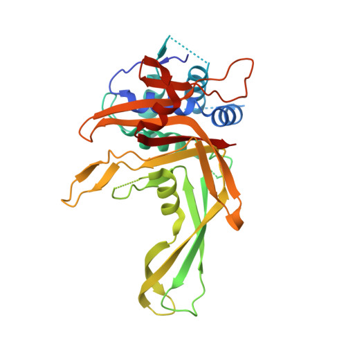
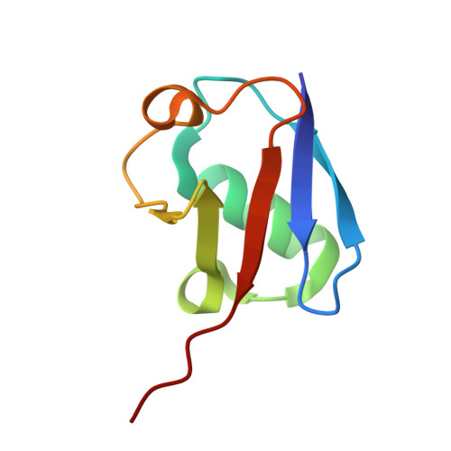
Chimeric deubiquitinase engineering reveals structural basis for specific inhibition of the mitophagy regulator USP30.

Kazi, N.H.Klink, N.Gallant, K.Kipka, G.M.Gersch, M.

(2025) Nat Struct Mol Biol 32: 1776-1786

  • DOI: https://doi.org/10.1038/s41594-025-01534-4
  • Primary Citation Related Structures: 
    9F19, 9F6G

  • PubMed Abstract: 

    The mitochondrial deubiquitinase ubiquitin-specific protease (USP) 30 negatively regulates PINK1-parkin-driven mitophagy. Whether enhanced mitochondrial quality control through inhibition of USP30 can protect dopaminergic neurons is currently being explored in a clinical trial for Parkinson's disease. However, the molecular basis for specific inhibition of USP30 by small molecules has remained elusive. Here we report the crystal structure of human USP30 in complex with a specific inhibitor, enabled by chimeric protein engineering. Our study uncovers how the inhibitor extends into a cryptic pocket facilitated by a compound-induced conformation of the USP30 switching loop. Our work underscores the potential of exploring induced pockets and conformational dynamics to obtain deubiquitinase inhibitors and identifies residues facilitating specific inhibition of USP30. More broadly, we delineate a conceptual framework for specific USP deubiquitinase inhibition based on a common ligandability hotspot in the Leu73 ubiquitin binding site and on diverse compound extensions. Collectively, our work establishes a generalizable chimeric protein-engineering strategy to aid deubiquitinase crystallization and enables structure-based drug design with relevance to neurodegeneration.


  • Organizational Affiliation
    • Chemical Genomics Center, Max Planck Institute of Molecular Physiology, Dortmund, Germany.

Macromolecule Content 

  • Total Structure Weight: 44.7 kDa 
  • Atom Count: 3,126 
  • Modeled Residue Count: 356 
  • Deposited Residue Count: 392 
  • Unique protein chains: 2

Macromolecules

Find similar proteins by:|  3D Structure
Entity ID: 1
MoleculeChains  Sequence LengthOrganismDetailsImage
Ubiquitin carboxyl-terminal hydrolase 30,Ubiquitin carboxyl-terminal hydrolase 14,Ubiquitin carboxyl-terminal hydrolase 35317Homo sapiensMutation(s): 1 
Gene Names: USP30USP14TGTUSP35KIAA1372USP34
EC: 3.4.19.12
UniProt & NIH Common Fund Data Resources
Find proteins for P54578 (Homo sapiens)
Explore P54578 
Go to UniProtKB:  P54578
PHAROS:  P54578
GTEx:  ENSG00000101557 
Find proteins for Q70CQ3 (Homo sapiens)
Explore Q70CQ3 
Go to UniProtKB:  Q70CQ3
PHAROS:  Q70CQ3
GTEx:  ENSG00000135093 
Find proteins for Q9P2H5 (Homo sapiens)
Explore Q9P2H5 
Go to UniProtKB:  Q9P2H5
PHAROS:  Q9P2H5
GTEx:  ENSG00000118369 
Entity Groups
Sequence Clusters30% Identity50% Identity70% Identity90% Identity95% Identity100% Identity
UniProt GroupsP54578Q70CQ3Q9P2H5
Sequence Annotations
Expand
Reference Sequence
Find similar proteins by:|  3D Structure
Entity ID: 2
MoleculeChains  Sequence LengthOrganismDetailsImage
Polyubiquitin-B75Homo sapiensMutation(s): 0 
Gene Names: UBB
UniProt & NIH Common Fund Data Resources
Find proteins for P0CG48 (Homo sapiens)
Explore P0CG48 
Go to UniProtKB:  P0CG48
PHAROS:  P0CG48
GTEx:  ENSG00000150991 
Entity Groups
Sequence Clusters30% Identity50% Identity70% Identity90% Identity95% Identity100% Identity
UniProt GroupP0CG48
Sequence Annotations
Expand
Reference Sequence

Small Molecules

Ligands 1 Unique
IDChains Name / Formula / InChI Key2D Diagram3D Interactions
AYE
(Subject of Investigation/LOI)

Query on AYE


Download:Ideal Coordinates CCD File
C [auth A]prop-2-en-1-amine
C3 H7 N
VVJKKWFAADXIJK-UHFFFAOYSA-N

Experimental Data & Validation

Experimental Data

  • Method: X-RAY DIFFRACTION
  • Resolution: 1.50 Å
  • R-Value Free:  0.211 (Depositor), 0.211 (DCC) 
  • R-Value Work:  0.183 (Depositor), 0.183 (DCC) 
  • R-Value Observed: 0.184 (Depositor) 
Space Group: P 21 21 21
Unit Cell:
Length ( Å )Angle ( ˚ )
a = 46.639α = 90
b = 50.706β = 90
c = 156.56γ = 90
Software Package:
Software NamePurpose
PHENIXrefinement
DIALSdata reduction
xia2data reduction
Aimlessdata scaling
PHASERphasing
Cootmodel building

Structure Validation

View Full Validation Report


Ligand Structure Quality Assessment 


Entry History 

& Funding Information

Deposition Data


Funding OrganizationLocationGrant Number
Max Planck SocietyGermanyCGCIII-352S

Revision History  (Full details and data files)

  • Version 1.0: 2025-03-19
    Type: Initial release
  • Version 1.1: 2025-05-14
    Changes: Database references
  • Version 1.2: 2025-10-01
    Changes: Database references