9EYQ | pdb_00009eyq

The structure of nonameric pore of RN1 variant of actinoporin Fav


Experimental Data Snapshot

  • Method: ELECTRON MICROSCOPY
  • Resolution: 3.28 Å
  • Aggregation State: PARTICLE 
  • Reconstruction Method: SINGLE PARTICLE 

Starting Model: experimental
View more details

wwPDB Validation 3D Report Full Report

Validation slider image for 9EYQ

This is version 1.1 of the entry. See complete history

Literature

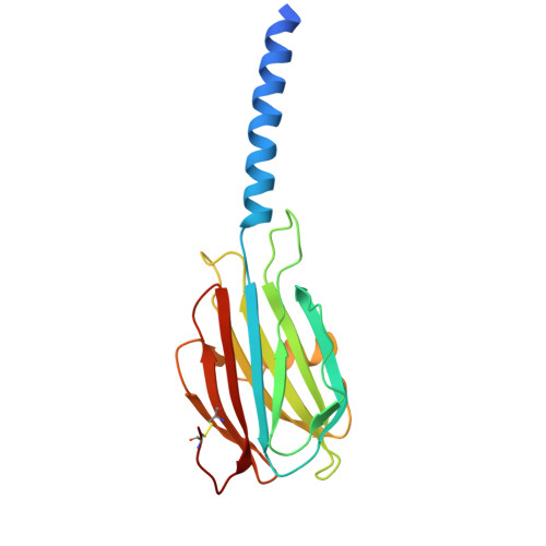
High-Throughput Human Histone Detection by an Engineered Actinoporin Nanopore.

Srnko, M.Solinc, G.Crnkovic, A.Merzel, F.Jordan, M.Wallace, E.J.Podobnik, M.Anderluh, G.

(2026) ACS Sens 11: 2016-2029

  • DOI: https://doi.org/10.1021/acssensors.5c03533
  • Primary Citation Related Structures: 
    9EYP, 9EYQ

  • PubMed Abstract: 

    The use of protein nanopores has proven to be very promising for the identification of proteins or the sequencing of peptides. However, their widespread use in biosensor applications is limited by more complex molecular properties of proteins in comparison to nucleic acids. Here, we developed an α-helical Fav nanopore from the coral Orbicella faveolata to detect human histones using a commercial MinION device. The engineered nanopores showed stable insertion into the polymeric membrane support. Bulk analysis of several tens to hundreds of pores per measurement revealed significant differences in mean current passing the pore and current noise resulting from capturing different full-length human histones or their post-translationally modified variants into the pore lumen. Importantly, detailed blocking analyses confirmed histone-specific signals that allow further discrimination between two medically important extracellular histones in mixtures. The newly developed Fav nanopore and high-throughput approach for the detection of biomedically important proteins is an important step towards the widespread use of nanopores in modern analytics.


  • Organizational Affiliation
    • Department of Molecular Biology and Nanobiotechnology, National Institute of Chemistry, Ljubljana 1000, Slovenia.

Macromolecule Content 

  • Total Structure Weight: 224.71 kDa 
  • Atom Count: 13,815 
  • Modeled Residue Count: 1,629 
  • Deposited Residue Count: 1,737 
  • Unique protein chains: 1

Macromolecules

Find similar proteins by:|  3D Structure
Entity ID: 1
MoleculeChains  Sequence LengthOrganismDetailsImage
Actinoporin
A, B, C, D, E
A, B, C, D, E, F, G, H, I
193Orbicella faveolataMutation(s): 0 

Small Molecules

Ligands 1 Unique
IDChains Name / Formula / InChI Key2D Diagram3D Interactions
A1H8M

Query on A1H8M


Download:Ideal Coordinates CCD File
AA [auth D]
AB [auth I]
BA [auth D]
BB [auth I]
CA [auth D]
AA [auth D],
AB [auth I],
BA [auth D],
BB [auth I],
CA [auth D],
DA [auth E],
EA [auth E],
FA [auth E],
GA [auth E],
HA [auth E],
IA [auth F],
J [auth A],
JA [auth F],
K [auth A],
KA [auth F],
L [auth A],
LA [auth F],
M [auth A],
MA [auth F],
N [auth A],
NA [auth G],
O [auth B],
OA [auth G],
P [auth B],
PA [auth G],
Q [auth B],
QA [auth G],
R [auth B],
RA [auth G],
S [auth B],
SA [auth H],
T [auth C],
TA [auth H],
U [auth C],
UA [auth H],
V [auth C],
VA [auth H],
W [auth C],
WA [auth H],
X [auth C],
XA [auth I],
Y [auth D],
YA [auth I],
Z [auth D],
ZA [auth I]
Sphingomyelin C18
C41 H84 N2 O6 P
LKQLRGMMMAHREN-YJFXYUILSA-O

Experimental Data & Validation

Experimental Data

  • Method: ELECTRON MICROSCOPY
  • Resolution: 3.28 Å
  • Aggregation State: PARTICLE 
  • Reconstruction Method: SINGLE PARTICLE 
EM Software:
TaskSoftware PackageVersion
MODEL REFINEMENTCoot0.98
MODEL REFINEMENTPHENIX1.19
MODEL REFINEMENTISOLDE1.6
RECONSTRUCTIONcryoSPARCv4

Structure Validation

View Full Validation Report


Entry History 

& Funding Information

Deposition Data


Funding OrganizationLocationGrant Number
Slovenian Research AgencySloveniaJ4-8225
Slovenian Research AgencySloveniaP1-0391
Other private--

Revision History  (Full details and data files)

  • Version 1.0: 2025-04-23
    Type: Initial release
  • Version 1.1: 2026-05-27
    Changes: Data collection, Database references