9ERZ | pdb_00009erz

Structure of CBL-TKBD bound to Ubiquitin-fused CBLock peptide

  • Classification: LIGASE
  • Organism(s): Homo sapiens
  • Expression System: Homo sapiens
  • Mutation(s): No 

  • Deposited: 2024-03-25 Released: 2025-05-14 
  • Deposition Author(s): Ahmed, S.F., Huang, D.T.
  • Funding Organization(s): Cancer Research UK, European Research Council (ERC)

Experimental Data Snapshot

  • Method: X-RAY DIFFRACTION
  • Resolution: 2.02 Å
  • R-Value Free: 
    0.229 (Depositor), 0.234 (DCC) 
  • R-Value Work: 
    0.178 (Depositor), 0.185 (DCC) 
  • R-Value Observed: 
    0.181 (Depositor) 

Starting Models: experimental
View more details

wwPDB Validation   3D Report Full Report


This is version 1.1 of the entry. See complete history


Literature

Locking CBL TKBD in its native conformation presents a novel therapeutic opportunity in mutant CBL-dependent leukemia.

Ahmed, S.F.Anand, J.Zhang, W.Buetow, L.Rishi, L.Mitchell, L.Bohlen, J.Lilla, S.Sibbet, G.J.Nixon, C.Patel, A.Majorek, K.A.Zanivan, S.Bustamante, J.C.Sidhu, S.S.Blyth, K.Huang, D.T.

(2025) Mol Ther 33: 3624-3643

  • DOI: https://doi.org/10.1016/j.ymthe.2025.04.042
  • Primary Citation of Related Structures:  
    9ERZ

  • PubMed Abstract: 

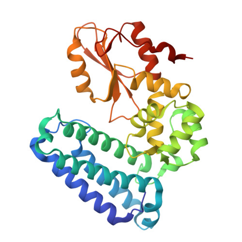
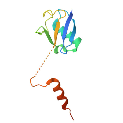
    Casitas B-lineage lymphoma (CBL) is an E3 ubiquitin ligase critical for negatively regulating receptor protein tyrosine kinases (RTKs). Deleterious CBL mutants lose E3 activity, but act as adaptors that gain function to cause myeloproliferative neoplasms. Currently, there is no targeted treatment available for patients with CBL mutant-dependent disorders. By combining phage-display technology and structure-based optimization, we discovered CBLock, a nanomolar affinity peptide inhibitor, that binds the substrate-binding site of CBL's tyrosine kinase binding domain (TKBD). CBLock disrupts the interaction between CBL mutants and RTKs, thereby impairing RTK-mediated priming of adaptor function of CBL mutants and downstream signaling. Notably, CBLock binds TKBD without inducing conformational changes, thereby preserving its ligand-free native conformation. In contrast, when CBL binds RTK substrates, TKBD undergoes a conformational change. Maintaining the native CBL TKBD conformation was crucial for CBLock to inhibit proliferation, induce cell cycle arrest, and promote apoptosis in leukemia cells harboring CBL mutations. In a mouse xenograft model of acute myeloid leukemia (AML), CBLock reduced tumor burden and improved survival rate. Moreover, CBLock inhibited the proliferation of cells derived from patients with CBL mutations. Therefore, inhibiting CBL TKBD in its native state presents a promising therapeutic opportunity in targeting mutant CBL-dependent leukemia.


  • Organizational Affiliation
    • Cancer Research UK Scotland Institute, Garscube Estate, Switchback Road, Glasgow, G61 1BD, United Kingdom.

Macromolecules
Find similar proteins by:  (by identity cutoff)  |  3D Structure
Entity ID: 1
MoleculeChains Sequence LengthOrganismDetailsImage
E3 ubiquitin-protein ligase CBL
A, C
311Homo sapiensMutation(s): 0 
Gene Names: CBLCBL2RNF55
EC: 2.3.2.27
UniProt & NIH Common Fund Data Resources
Find proteins for P22681 (Homo sapiens)
Explore P22681 
Go to UniProtKB:  P22681
PHAROS:  P22681
GTEx:  ENSG00000110395 
Entity Groups  
Sequence Clusters30% Identity50% Identity70% Identity90% Identity95% Identity100% Identity
UniProt GroupP22681
Sequence Annotations
Expand
  • Reference Sequence
Find similar proteins by:  (by identity cutoff)  |  3D Structure
Entity ID: 2
MoleculeChains Sequence LengthOrganismDetailsImage
Polyubiquitin-C,Ub-fused CBLock peptide
B, D
107Homo sapiensMutation(s): 0 
Gene Names: UBC
UniProt & NIH Common Fund Data Resources
Find proteins for P0CG48 (Homo sapiens)
Explore P0CG48 
Go to UniProtKB:  P0CG48
PHAROS:  P0CG48
GTEx:  ENSG00000150991 
Entity Groups  
Sequence Clusters30% Identity50% Identity70% Identity90% Identity95% Identity100% Identity
UniProt GroupP0CG48
Sequence Annotations
Expand
  • Reference Sequence
Experimental Data & Validation

Experimental Data

  • Method: X-RAY DIFFRACTION
  • Resolution: 2.02 Å
  • R-Value Free:  0.229 (Depositor), 0.234 (DCC) 
  • R-Value Work:  0.178 (Depositor), 0.185 (DCC) 
  • R-Value Observed: 0.181 (Depositor) 
Space Group: P 1 21 1
Unit Cell:
Length ( Å )Angle ( ˚ )
a = 59.783α = 90
b = 87.324β = 99.39
c = 84.353γ = 90
Software Package:
Software NamePurpose
PHENIXrefinement
XDSdata reduction
xia2data scaling
PHASERphasing

Structure Validation

View Full Validation Report


Entry History & Funding Information

Deposition Data


Funding OrganizationLocationGrant Number
Cancer Research UKUnited KingdomA23278
European Research Council (ERC)European Union647849

Revision History  (Full details and data files)

  • Version 1.0: 2025-05-14
    Type: Initial release
  • Version 1.1: 2025-08-20
    Changes: Database references