9ERY | pdb_00009ery

Co-crystal strucutre of PD-L1 with low molecular weight inhibitor


Experimental Data Snapshot

  • Method: X-RAY DIFFRACTION
  • Resolution: 2.70 Å
  • R-Value Free: 
    0.276 (Depositor), 0.275 (DCC) 
  • R-Value Work: 
    0.222 (Depositor), 0.221 (DCC) 

Starting Model: experimental
View more details

wwPDB Validation 3D Report Full Report

Validation slider image for 9ERY

Ligand Structure Quality Assessment 


This is version 1.2 of the entry. See complete history

Literature

Design, synthesis, and evaluation of antitumor activity of 2-arylmethoxy-4-(2-fluoromethyl-biphenyl-3-ylmethoxy) benzylamine derivatives as PD-1/PD-l1 inhibitors.

Zhang, F.Zhang, H.Zhou, S.Plewka, J.Wang, M.Sun, S.Wu, C.Yu, Q.Zhu, M.Awadasseid, A.Wu, Y.Magiera-Mularz, K.Zhang, W.

(2024) Eur J Med Chem 276: 116683-116683

  • DOI: https://doi.org/10.1016/j.ejmech.2024.116683
  • Primary Citation Related Structures: 
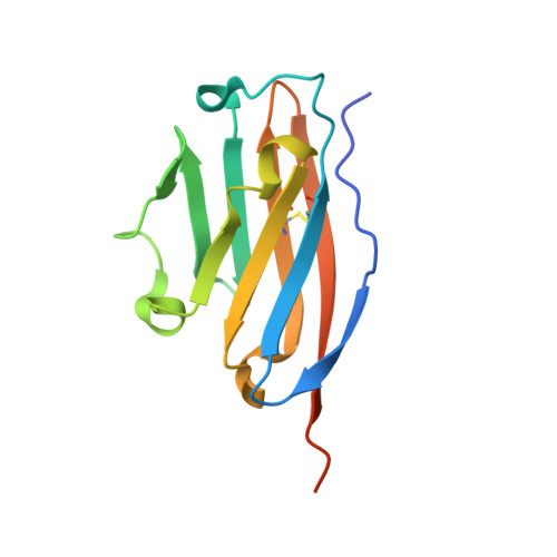
    9ERY

  • PubMed Abstract: 

    A series of novel 2-arylmethoxy-4-(2-fluoromethyl-biphenyl-3-ylmethoxy) benzylamine derivatives was designed, synthesized, and evaluated for their antitumor effects as PD-1/PD-L1 inhibitors both in vitro and in vivo. Firstly, the ability of these compounds to block the PD-1/PD-L1 immune checkpoint was assessed using the homogeneous time-resolved fluorescence (HTRF) assay. Two of the compounds can strongly block the PD-1/PD-L1 interaction, with IC 50 values of less than 10 nM, notably, compound HD10 exhibited significant clinical potential by inhibiting the PD-1/PD-L1 interaction with an IC 50 value of 3.1 nM. Further microscale thermophoresis (MST) analysis demonstrated that HD10 had strong interaction with PD-L1 protein. Co-crystal structure (2.7 Å) analysis of HD10 in complex with the PD-L1 protein revealed a strong affinity between the compound and the target PD-L1 dimer. This provides a solid theoretical basis for further in vitro and in vivo studies. Next, a typical cell-based experiment demonstrated that HD10 could remarkably prevent the interaction of hPD-1 293 T cells from human recombinant PD-L1 protein, effectively restoring T cell function, and promoting IFN-γ secretion in a dose-dependent manner. Moreover, HD10 was effective in suppressing tumor growth (TGI = 57.31 %) in a PD-1/PD-L1 humanized mouse model without obvious toxicity. Flow cytometry, qPCR, and immunohistochemistry data suggested that HD10 inhibits tumor growth by activating the immune system in vivo. Based on these results, it seems likely that HD10 is a promising clinical candidate that should be further investigated.


  • Organizational Affiliation
    • Lab of Chemical Biology and Molecular Drug Design, College of Pharmaceutical Science, Zhejiang University of Technology, Deqing, 313299, China; Institute of Drug Development & Chemical Biology, Zhejiang University of Technology, Deqing, 313299, China.

Macromolecule Content 

  • Total Structure Weight: 30.58 kDa 
  • Atom Count: 1,889 
  • Modeled Residue Count: 232 
  • Deposited Residue Count: 262 
  • Unique protein chains: 1

Macromolecules

Find similar proteins by:|  3D Structure
Entity ID: 1
MoleculeChains  Sequence LengthOrganismDetailsImage
Programmed cell death 1 ligand 1
A, B
131Homo sapiensMutation(s): 0 
Gene Names: CD274B7H1PDCD1L1PDCD1LG1PDL1
UniProt & NIH Common Fund Data Resources
Find proteins for Q9NZQ7 (Homo sapiens)
Explore Q9NZQ7 
Go to UniProtKB:  Q9NZQ7
PHAROS:  Q9NZQ7
GTEx:  ENSG00000120217 
Entity Groups
Sequence Clusters30% Identity50% Identity70% Identity90% Identity95% Identity100% Identity
UniProt GroupQ9NZQ7
Sequence Annotations
Expand
Reference Sequence

Small Molecules

Ligands 2 Unique
IDChains Name / Formula / InChI Key2D Diagram3D Interactions
VVH
(Subject of Investigation/LOI)

Query on VVH


Download:Ideal Coordinates CCD File
C [auth A]5-[[5-[[2-[bis(fluoranyl)methyl]-3-(2,3-dihydro-1,4-benzodioxin-6-yl)phenyl]methoxy]-2-[(2-hydroxyethylamino)methyl]phenoxy]methyl]pyridine-3-carbonitrile
C32 H29 F2 N3 O5
YNAHCBRGGPSNDZ-UHFFFAOYSA-N
SO4

Query on SO4


Download:Ideal Coordinates CCD File
D [auth A]SULFATE ION
O4 S
QAOWNCQODCNURD-UHFFFAOYSA-L

Experimental Data & Validation

Experimental Data

  • Method: X-RAY DIFFRACTION
  • Resolution: 2.70 Å
  • R-Value Free:  0.276 (Depositor), 0.275 (DCC) 
  • R-Value Work:  0.222 (Depositor), 0.221 (DCC) 
Space Group: P 31 2 1
Unit Cell:
Length ( Å )Angle ( ˚ )
a = 74.534α = 90
b = 74.534β = 90
c = 95.392γ = 120
Software Package:
Software NamePurpose
REFMACrefinement
PDB-REDOrefinement
Aimlessdata scaling
PHASERphasing
XDSdata reduction

Structure Validation

View Full Validation Report


Ligand Structure Quality Assessment 


Entry History 

& Funding Information

Deposition Data


Funding OrganizationLocationGrant Number
National Natural Science Foundation of China (NSFC)China21877101
National Natural Science Foundation of China (NSFC)China22177105

Revision History  (Full details and data files)

  • Version 1.0: 2024-07-24
    Type: Initial release
  • Version 1.1: 2024-07-31
    Changes: Database references
  • Version 1.2: 2024-10-23
    Changes: Structure summary