9EQF | pdb_00009eqf

Crystal structure of the L-arginine hydroxylase VioC MeHis316, bound to Fe(II), L-arginine, and succinate

  • Classification: OXIDOREDUCTASE
  • Organism(s): synthetic construct
  • Expression System: Escherichia coli
  • Mutation(s): No 

  • Deposited: 2024-03-21 Released: 2024-07-31 
  • Deposition Author(s): Hardy, F.J.
  • Funding Organization(s): European Research Council (ERC), Biotechnology and Biological Sciences Research Council (BBSRC), Engineering and Physical Sciences Research Council

Experimental Data Snapshot

  • Method: X-RAY DIFFRACTION
  • Resolution: 1.60 Å
  • R-Value Free: 
    0.187 (Depositor), 0.186 (DCC) 
  • R-Value Work: 
    0.158 (Depositor), 0.157 (DCC) 
  • R-Value Observed: 
    0.159 (Depositor) 

Starting Model: experimental
View more details

wwPDB Validation 3D Report Full Report

Validation slider image for 9EQF

Ligand Structure Quality Assessment 


This is version 1.1 of the entry. See complete history

Literature

Probing Ferryl Reactivity in a Nonheme Iron Oxygenase Using an Expanded Genetic Code.

Hardy, F.J.Quesne, M.G.Gerard, E.F.Zhao, J.Ortmayer, M.Taylor, C.J.Ali, H.S.Slater, J.W.Levy, C.W.Heyes, D.J.Bollinger Jr., J.M.de Visser, S.P.Green, A.P.

(2024) ACS Catal 14: 11584-11590

  • DOI: https://doi.org/10.1021/acscatal.4c02365
  • Primary Citation Related Structures: 
    9EQF

  • PubMed Abstract: 

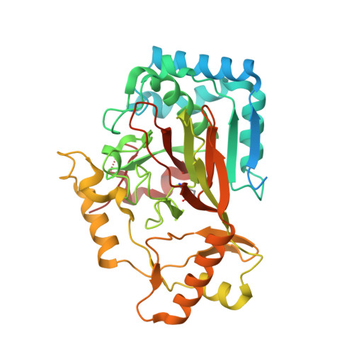
    The ability to introduce noncanonical amino acids as axial ligands in heme enzymes has provided a powerful experimental tool for studying the structure and reactivity of their Fe IV =O ("ferryl") intermediates. Here, we show that a similar approach can be used to perturb the conserved Fe coordination environment of 2-oxoglutarate (2OG) dependent oxygenases, a versatile class of enzymes that employ highly-reactive ferryl intermediates to mediate challenging C-H functionalizations. Replacement of one of the cis-disposed histidine ligands in the oxygenase VioC with a less electron donating N δ -methyl-histidine (MeHis) preserves both catalytic function and reaction selectivity. Significantly, the key ferryl intermediate responsible for C-H activation can be accumulated in both the wildtype and the modified protein. In contrast to heme enzymes, where metal-oxo reactivity is extremely sensitive to the nature of the proximal ligand, the rates of C-H activation and the observed large kinetic isotope effects are only minimally affected by axial ligand replacement in VioC. This study showcases a powerful tool for modulating the coordination sphere of nonheme iron enzymes that will enhance our understanding of the factors governing their divergent activities.


  • Organizational Affiliation
    • Department of Chemistry & Manchester Institute of Biotechnology, The University of Manchester, 131 Princess Street, Manchester M1 7DN, U.K.

Macromolecule Content 

  • Total Structure Weight: 43.84 kDa 
  • Atom Count: 3,187 
  • Modeled Residue Count: 335 
  • Deposited Residue Count: 394 
  • Unique protein chains: 1

Macromolecules

Find similar proteins by:|  3D Structure
Entity ID: 1
MoleculeChains  Sequence LengthOrganismDetailsImage
Alpha-ketoglutarate-dependent L-arginine hydroxylase394synthetic constructMutation(s): 0 
Gene Names: vioC
EC: 1.14.11.41
UniProt
Find proteins for Q6WZB0 (Streptomyces vinaceus)
Explore Q6WZB0 
Go to UniProtKB:  Q6WZB0
Entity Groups
Sequence Clusters30% Identity50% Identity70% Identity90% Identity95% Identity100% Identity
UniProt GroupQ6WZB0
Sequence Annotations
Expand
Reference Sequence

Small Molecules

Ligands 5 Unique
IDChains Name / Formula / InChI Key2D Diagram3D Interactions
ARG
(Subject of Investigation/LOI)

Query on ARG


Download:Ideal Coordinates CCD File
D [auth A]ARGININE
C6 H15 N4 O2
ODKSFYDXXFIFQN-BYPYZUCNSA-O
SIN
(Subject of Investigation/LOI)

Query on SIN


Download:Ideal Coordinates CCD File
C [auth A]SUCCINIC ACID
C4 H6 O4
KDYFGRWQOYBRFD-UHFFFAOYSA-N
PEG

Query on PEG


Download:Ideal Coordinates CCD File
F [auth A]DI(HYDROXYETHYL)ETHER
C4 H10 O3
MTHSVFCYNBDYFN-UHFFFAOYSA-N
EDO

Query on EDO


Download:Ideal Coordinates CCD File
E [auth A]1,2-ETHANEDIOL
C2 H6 O2
LYCAIKOWRPUZTN-UHFFFAOYSA-N
FE2
(Subject of Investigation/LOI)

Query on FE2


Download:Ideal Coordinates CCD File
B [auth A]FE (II) ION
Fe
CWYNVVGOOAEACU-UHFFFAOYSA-N
Modified Residues  1 Unique
IDChains TypeFormula2D DiagramParent
MHS
Query on MHS
A
L-PEPTIDE LINKINGC7 H11 N3 O2HIS

Experimental Data & Validation

Experimental Data

  • Method: X-RAY DIFFRACTION
  • Resolution: 1.60 Å
  • R-Value Free:  0.187 (Depositor), 0.186 (DCC) 
  • R-Value Work:  0.158 (Depositor), 0.157 (DCC) 
  • R-Value Observed: 0.159 (Depositor) 
Space Group: C 1 2 1
Unit Cell:
Length ( Å )Angle ( ˚ )
a = 81.145α = 90
b = 67.107β = 109.221
c = 62.945γ = 90
Software Package:
Software NamePurpose
PHENIXrefinement
DIALSdata reduction
DIALSdata scaling
PHENIXphasing

Structure Validation

View Full Validation Report


Ligand Structure Quality Assessment 


Entry History 

& Funding Information

Deposition Data

  • Released Date: 2024-07-31 
  • Deposition Author(s): Hardy, F.J.

Funding OrganizationLocationGrant Number
European Research Council (ERC)European Union757991
Biotechnology and Biological Sciences Research Council (BBSRC)United KingdomBB/M027023/1
Engineering and Physical Sciences Research CouncilUnited KingdomEP/W524347/1

Revision History  (Full details and data files)

  • Version 1.0: 2024-07-31
    Type: Initial release
  • Version 1.1: 2024-08-21
    Changes: Database references