9EOI | pdb_00009eoi

Crystal structure of the GH19 endolysin from Pseudomonas aeruginosa


Experimental Data Snapshot

  • Method: X-RAY DIFFRACTION
  • Resolution: 1.77 Å
  • R-Value Free: 
    0.254 (Depositor), 0.254 (DCC) 
  • R-Value Work: 
    0.216 (Depositor), 0.216 (DCC) 
  • R-Value Observed: 
    0.218 (Depositor) 

Starting Model: in silico
View more details

wwPDB Validation   3D Report Full Report


Ligand Structure Quality Assessment 


This is version 1.1 of the entry. See complete history


Literature

Pseudomonas aeruginosa Cryptic Prophage Endolysin Is a Highly Active Muramidase.

Thoren Edvardsen, P.K.Englund, A.N.Kjendseth Rohr, A.Mesnage, S.Vaaje-Kolstad, G.

(2025) Biochemistry 64: 3446-3458

  • DOI: https://doi.org/10.1021/acs.biochem.5c00142
  • Primary Citation of Related Structures:  
    9EOI

  • PubMed Abstract: 

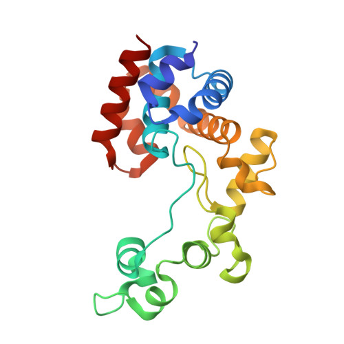
    Endolysins are phage-encoded enzymes that cleave the peptidoglycan of host bacteria. These enzymes have gained considerable attention due to their ability to cause cell lysis, making them candidates as antibacterial agents. Most Pseudomonas aeruginosa genomes, including the common laboratory strains PAO1 and UCBPP-PA14, contain a cryptic prophage encoding a glycoside hydrolase family 19 endolysin (named Pa GH19Lys in the present study). Family 19 glycoside hydrolases are known to target peptidoglycan and chitin-type substrates. Pa GH19Lys was not active toward chitin but exhibited activity toward chloroform-treated Gram-negative bacteria, displaying ∼10,000-fold higher activity than hen egg white lysozyme. Analysis of products derived from Pa GH19Lys activity toward purified P. aeruginosa peptidoglycan showed that the enzyme catalyzed hydrolysis of the β-1,4 linkage between N- acetylmuramic acid and N- acetyl-d-glucosamine, classifying the enzyme as a muramidase. Finally, the crystal structure of Pa GH19Lys was determined and solved to 1.8 Å resolution. The structure of the enzyme showed a globular α-helical fold possessing a deep but relatively open catalytic cleft.


  • Organizational Affiliation
    • Faculty of Chemistry, Biotechnology, and Food Science, Norwegian University of Life Sciences, Ås 1432, Norway.

Macromolecules
Find similar proteins by:  (by identity cutoff)  |  3D Structure
Entity ID: 1
MoleculeChains Sequence LengthOrganismDetailsImage
Putative lytic enzyme
A, B
209Pseudomonas aeruginosa UCBPP-PA14Mutation(s): 0 
Gene Names: lysPA14_08160
UniProt
Find proteins for G3XD75 (Pseudomonas aeruginosa (strain ATCC 15692 / DSM 22644 / CIP 104116 / JCM 14847 / LMG 12228 / 1C / PRS 101 / PAO1))
Explore G3XD75 
Go to UniProtKB:  G3XD75
Entity Groups  
Sequence Clusters30% Identity50% Identity70% Identity90% Identity95% Identity100% Identity
UniProt GroupG3XD75
Sequence Annotations
Expand
  • Reference Sequence
Experimental Data & Validation

Experimental Data

  • Method: X-RAY DIFFRACTION
  • Resolution: 1.77 Å
  • R-Value Free:  0.254 (Depositor), 0.254 (DCC) 
  • R-Value Work:  0.216 (Depositor), 0.216 (DCC) 
  • R-Value Observed: 0.218 (Depositor) 
Space Group: P 21 21 21
Unit Cell:
Length ( Å )Angle ( ˚ )
a = 65.406α = 90
b = 79.696β = 90
c = 91.615γ = 90
Software Package:
Software NamePurpose
PHENIXrefinement
PHENIXrefinement
XDSdata reduction
Aimlessdata scaling
PHENIXphasing

Structure Validation

View Full Validation Report


Ligand Structure Quality Assessment 


Entry History & Funding Information

Deposition Data


Funding OrganizationLocationGrant Number
Other governmentNorway--

Revision History  (Full details and data files)

  • Version 1.0: 2025-02-19
    Type: Initial release
  • Version 1.1: 2025-09-03
    Changes: Database references