9EI9 | pdb_00009ei9

Cryo-EM structure of 5E10 Fab in complex with H3 influenza Victoria 2011 HA trimer


Experimental Data Snapshot

  • Method: ELECTRON MICROSCOPY
  • Resolution: 3.89 Å
  • Aggregation State: PARTICLE 
  • Reconstruction Method: SINGLE PARTICLE 

Starting Model: in silico
View more details

wwPDB Validation   3D Report Full Report


This is version 1.2 of the entry. See complete history


Literature

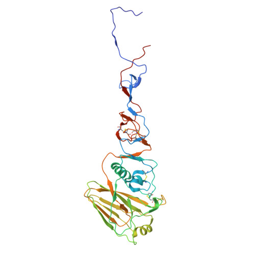
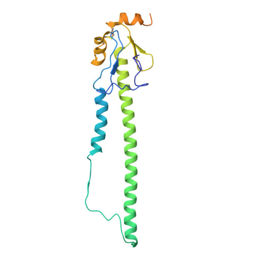
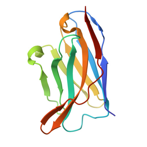
The N terminus of H3-influenza hemagglutinin as a site-of-vulnerability to neutralizing antibody.

Rawi, R.Morano, N.C.Cheung, C.S.Du, H.Gorman, J.Prabhakaran, M.Becker, J.E.Bylund, T.Charaf, S.Chen, X.Lee, M.Harris, D.R.Olia, A.S.Ou, L.Wang, L.Wang, S.Zhang, B.Kanekiyo, M.McDermott, A.B.Zhou, T.Shapiro, L.Kwong, P.D.

(2025) Structure 33: 1820

  • DOI: https://doi.org/10.1016/j.str.2025.07.015
  • Primary Citation of Related Structures:  
    9EI8, 9EI9

  • PubMed Abstract: 

    The N terminus of the H3 subtype of influenza virus hemagglutinin is ∼10 residues longer than the N termini of most other hemagglutinins. As conserved, exposed, and linear regions may be good vaccine targets, we investigated the vaccine utility of the extended H3-N terminus. First, we identified antibody 5E10, for which structure and binding analyses revealed recognition of the H3-N terminus. Second, we immunized mice with immunogens incorporating the H3-N terminus, boosted with hemagglutinin trimer, and isolated antibodies from immunogen-elicited B cells that bound both H3-N terminus and hemagglutinin trimer. However, hemagglutinin-complex structures of two such antibodies, 3864-6 and 3864-10, that neutralized H3-influenza strains, revealed only peripheral recognition of the hemagglutinin N terminus. Collectively, these results reveal the N terminus of H3 hemagglutinin to be a suboptimal vaccine target and suggest that-in addition to being conserved, flexible, and accessible-other factors influence the elicitation of potent broadly neutralizing responses.


  • Organizational Affiliation
    • Vaccine Research Center, National Institute of Allergy and Infectious Diseases, National Institutes of Health, Bethesda, MD 20892, USA. Electronic address: reda.rawi@nih.gov.

Macromolecules
Find similar proteins by:  (by identity cutoff)  |  3D Structure
Entity ID: 1
MoleculeChains Sequence LengthOrganismDetailsImage
Hemagglutinin HA1A [auth B],
B [auth C],
E [auth G]
334Influenza A virus (A/Victoria/361/2011(H3N2))Mutation(s): 0 
Gene Names: HA
UniProt
Find proteins for A0A5P1MU07 (Influenza A virus)
Explore A0A5P1MU07 
Go to UniProtKB:  A0A5P1MU07
Entity Groups  
Sequence Clusters30% Identity50% Identity70% Identity90% Identity95% Identity100% Identity
UniProt GroupA0A5P1MU07
Glycosylation
Glycosylation Sites: 8
Sequence Annotations
Expand
  • Reference Sequence
Find similar proteins by:  (by identity cutoff)  |  3D Structure
Entity ID: 2
MoleculeChains Sequence LengthOrganismDetailsImage
Hemagglutinin HA2C [auth E],
D [auth F],
F [auth I]
222Influenza A virus (A/Victoria/361/2011(H3N2))Mutation(s): 0 
Gene Names: HA
UniProt
Find proteins for A0A3G2CC79 (Influenza A virus)
Explore A0A3G2CC79 
Go to UniProtKB:  A0A3G2CC79
Entity Groups  
Sequence Clusters30% Identity50% Identity70% Identity90% Identity95% Identity100% Identity
UniProt GroupA0A3G2CC79
Glycosylation
Glycosylation Sites: 1
Sequence Annotations
Expand
  • Reference Sequence
Find similar proteins by:  (by identity cutoff)  |  3D Structure
Entity ID: 3
MoleculeChains Sequence LengthOrganismDetailsImage
5E10 Fab Heavy chainG [auth A],
I [auth H]
123Mus musculusMutation(s): 0 
Entity Groups  
Sequence Clusters30% Identity50% Identity70% Identity90% Identity95% Identity100% Identity
Sequence Annotations
Expand
  • Reference Sequence
Find similar proteins by:  (by identity cutoff)  |  3D Structure
Entity ID: 4
MoleculeChains Sequence LengthOrganismDetailsImage
5E10 Fab Light chainH [auth D],
J
111Mus musculusMutation(s): 0 
Entity Groups  
Sequence Clusters30% Identity50% Identity70% Identity90% Identity95% Identity100% Identity
Sequence Annotations
Expand
  • Reference Sequence
Oligosaccharides

Help

Entity ID: 5
MoleculeChains Length2D Diagram Glycosylation3D Interactions
2-acetamido-2-deoxy-beta-D-glucopyranose-(1-4)-2-acetamido-2-deoxy-beta-D-glucopyranose
K, M, N, P
2N-Glycosylation
Glycosylation Resources
GlyTouCan:  G42666HT
GlyCosmos:  G42666HT
GlyGen:  G42666HT
Entity ID: 6
MoleculeChains Length2D Diagram Glycosylation3D Interactions
beta-D-mannopyranose-(1-4)-2-acetamido-2-deoxy-beta-D-glucopyranose-(1-4)-2-acetamido-2-deoxy-beta-D-glucopyranose
L, O, Q
3N-Glycosylation
Glycosylation Resources
GlyTouCan:  G15407YE
GlyCosmos:  G15407YE
GlyGen:  G15407YE
Small Molecules
Ligands 1 Unique
IDChains Name / Formula / InChI Key2D Diagram3D Interactions
NAG
Query on NAG

Download Ideal Coordinates CCD File 
AA [auth C]
BA [auth C]
CA [auth E]
DA [auth F]
EA [auth G]
AA [auth C],
BA [auth C],
CA [auth E],
DA [auth F],
EA [auth G],
FA [auth G],
GA [auth G],
HA [auth G],
IA [auth G],
JA [auth G],
KA [auth I],
R [auth B],
S [auth B],
T [auth B],
U [auth B],
V [auth B],
W [auth C],
X [auth C],
Y [auth C],
Z [auth C]
2-acetamido-2-deoxy-beta-D-glucopyranose
C8 H15 N O6
OVRNDRQMDRJTHS-FMDGEEDCSA-N
Experimental Data & Validation

Experimental Data

  • Method: ELECTRON MICROSCOPY
  • Resolution: 3.89 Å
  • Aggregation State: PARTICLE 
  • Reconstruction Method: SINGLE PARTICLE 
EM Software:
TaskSoftware PackageVersion
RECONSTRUCTIONcryoSPARC3
MODEL REFINEMENTPHENIX

Structure Validation

View Full Validation Report


Entry History & Funding Information

Deposition Data


Funding OrganizationLocationGrant Number
National Institutes of Health/National Institute of General Medical Sciences (NIH/NIGMS)United StatesGM103310
Simons FoundationUnited StatesSF349247

Revision History  (Full details and data files)

  • Version 1.0: 2025-07-16
    Type: Initial release
  • Version 1.1: 2025-08-27
    Changes: Data collection, Database references
  • Version 1.2: 2025-11-19
    Changes: Data collection, Database references