9EHI | pdb_00009ehi

Crystal structure of ENL YEATS in complex with histone H3 methacrylated at K18


Experimental Data Snapshot

  • Method: X-RAY DIFFRACTION
  • Resolution: 2.60 Å
  • R-Value Free: 
    0.267 (Depositor), 0.268 (DCC) 
  • R-Value Work: 
    0.219 (Depositor), 0.225 (DCC) 
  • R-Value Observed: 
    0.222 (Depositor) 

Starting Model: experimental
View more details

wwPDB Validation   3D Report Full Report


This is version 1.2 of the entry. See complete history


Literature

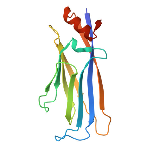
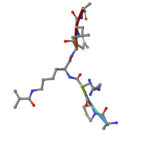
The YEATS domain is a selective reader of histone methacrylation.

Becht, D.C.Song, J.Selvam, K.Yin, K.Bai, W.Zhao, Y.Wu, R.Zheng, Y.G.Kutateladze, T.G.

(2025) Structure 33: 1233-1239.e5

  • DOI: https://doi.org/10.1016/j.str.2025.04.010
  • Primary Citation of Related Structures:  
    9EHI

  • PubMed Abstract: 

    Metabolically regulated lysine acylation modifications in proteins play a major role in epigenetic processes and cellular homeostasis. A new type of histone acylation, lysine methacrylation, has recently been identified but remains poorly characterized. Here, we show that lysine methacrylation can be generated through metabolism of sodium methacrylate and enzymatically removed in cells, and that the YEATS domain but not bromodomain recognizes this modification. Structural and biochemical analyses reveal the π-π-π-stacking mechanism for binding of the YEATS domain of ENL to methacrylated histone H3K18 (H3K18mc). Using mass spectrometry proteomics, we demonstrate that methacrylate induces global methacrylation of a set of proteins that differs from the set of methacrylated proteins associated with valine metabolism. These findings suggest that high levels of methacrylate may potentially perturb cellular functions of these proteins by altering protein methacrylation profiles.


  • Organizational Affiliation
    • Department of Pharmacology, University of Colorado School of Medicine, Aurora, CO 80045, USA.

Macromolecules
Find similar proteins by:  (by identity cutoff)  |  3D Structure
Entity ID: 1
MoleculeChains Sequence LengthOrganismDetailsImage
Protein ENL
A, B, C, D, E
A, B, C, D, E, F, G, H, I, J, K, L, M, N, O, P
145Homo sapiensMutation(s): 0 
Gene Names: MLLT1ENLLTG19YEATS1
UniProt & NIH Common Fund Data Resources
Find proteins for Q03111 (Homo sapiens)
Explore Q03111 
Go to UniProtKB:  Q03111
PHAROS:  Q03111
GTEx:  ENSG00000130382 
Entity Groups  
Sequence Clusters30% Identity50% Identity70% Identity90% Identity95% Identity100% Identity
UniProt GroupQ03111
Sequence Annotations
Expand
  • Reference Sequence

Find similar proteins by:  Sequence   |   3D Structure  

Entity ID: 2
MoleculeChains Sequence LengthOrganismDetailsImage
Histone H3.17Homo sapiensMutation(s): 0 
UniProt & NIH Common Fund Data Resources
Find proteins for P68431 (Homo sapiens)
Explore P68431 
Go to UniProtKB:  P68431
PHAROS:  P68431
Entity Groups  
UniProt GroupP68431
Sequence Annotations
Expand
  • Reference Sequence
Experimental Data & Validation

Experimental Data

  • Method: X-RAY DIFFRACTION
  • Resolution: 2.60 Å
  • R-Value Free:  0.267 (Depositor), 0.268 (DCC) 
  • R-Value Work:  0.219 (Depositor), 0.225 (DCC) 
  • R-Value Observed: 0.222 (Depositor) 
Space Group: P 1
Unit Cell:
Length ( Å )Angle ( ˚ )
a = 53.463α = 87.59
b = 98.413β = 90
c = 118.449γ = 90.02
Software Package:
Software NamePurpose
REFMACrefinement
Aimlessdata scaling
XDSdata reduction
PHASERphasing

Structure Validation

View Full Validation Report


Entry History & Funding Information

Deposition Data


Funding OrganizationLocationGrant Number
Not funded--

Revision History  (Full details and data files)

  • Version 1.0: 2025-04-09
    Type: Initial release
  • Version 1.1: 2025-06-11
    Changes: Database references
  • Version 1.2: 2025-07-16
    Changes: Database references