
 Download Mendeley
Crystal structure of Cryptosporidium parvum N-myristoyltransferase with bound myristoyl-CoA and inhibitor 20045
Staker, B.L., Fan, E.To be published.
Experimental Data Snapshot
Starting Model: experimental
View more details
Entity ID: 1 | |||||
|---|---|---|---|---|---|
| Molecule | Chains | Sequence Length | Organism | Details | Image |
| Glycylpeptide N-tetradecanoyltransferase | 431 | Cryptosporidium parvum Iowa II | Mutation(s): 0 Gene Names: cgd3_320 EC: 2.3.1.97 |  | |
UniProt | |||||
Entity Groups | |||||
| Sequence Clusters | 30% Identity50% Identity70% Identity90% Identity95% Identity100% Identity | ||||
| UniProt Group | Q5CV46 | ||||
Sequence AnnotationsExpand | |||||
Reference Sequence | |||||
| Ligands 6 Unique | |||||
|---|---|---|---|---|---|
| ID | Chains | Name / Formula / InChI Key | 2D Diagram | 3D Interactions | |
| MYA (Subject of Investigation/LOI) Download:Ideal Coordinates CCD File | C [auth A], J [auth B] | TETRADECANOYL-COA C35 H62 N7 O17 P3 S DUAFKXOFBZQTQE-QSGBVPJFSA-N | 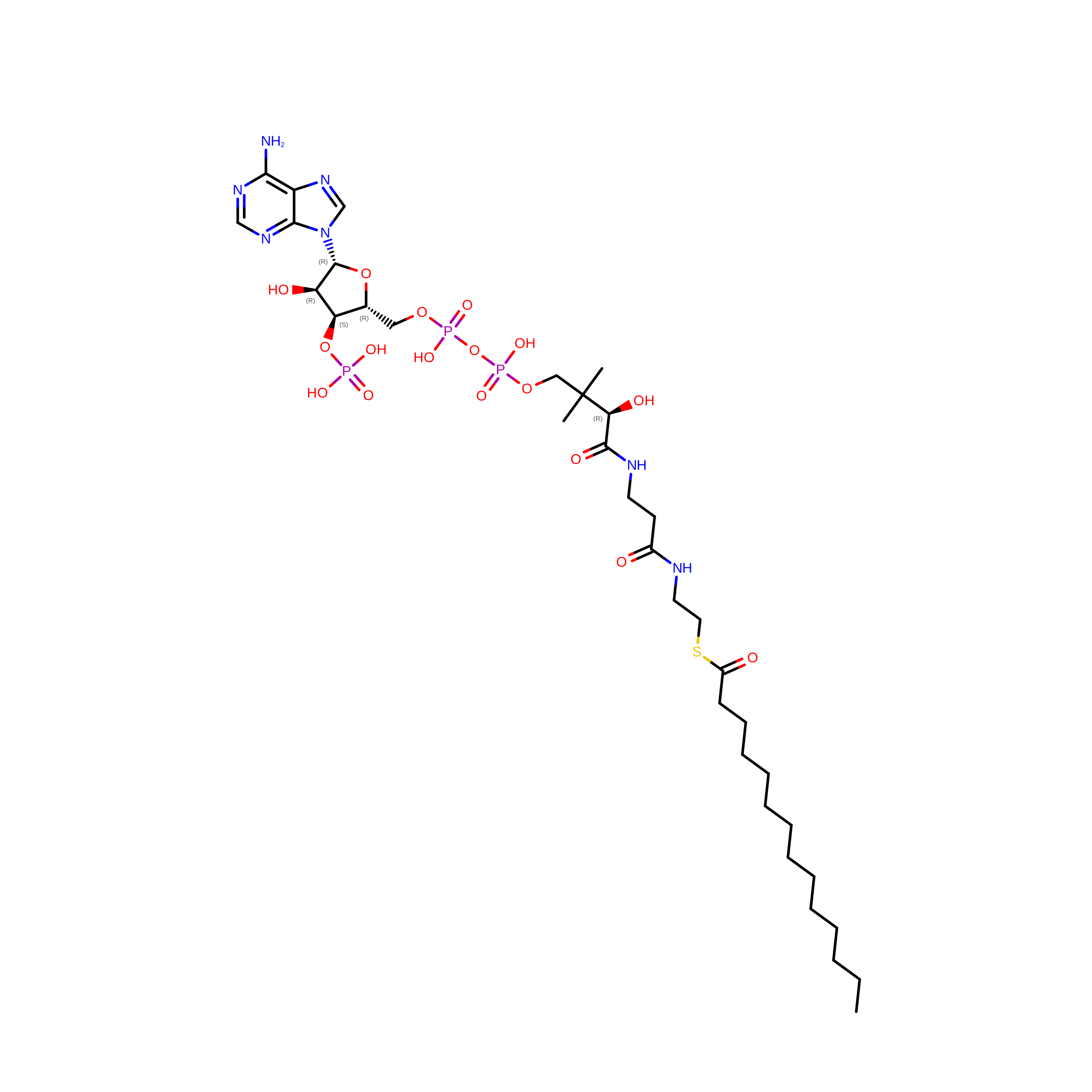 | ||
| A1BHP (Subject of Investigation/LOI) Download:Ideal Coordinates CCD File | D [auth A] | (2M)-N-(4-methyl-1,3-thiazol-2-yl)-2-[2-(piperazin-1-yl)phenyl]-1H-1,3-benzimidazole-4-carboxamide C22 H22 N6 O S XFFHSVKASCGESU-UHFFFAOYSA-N | 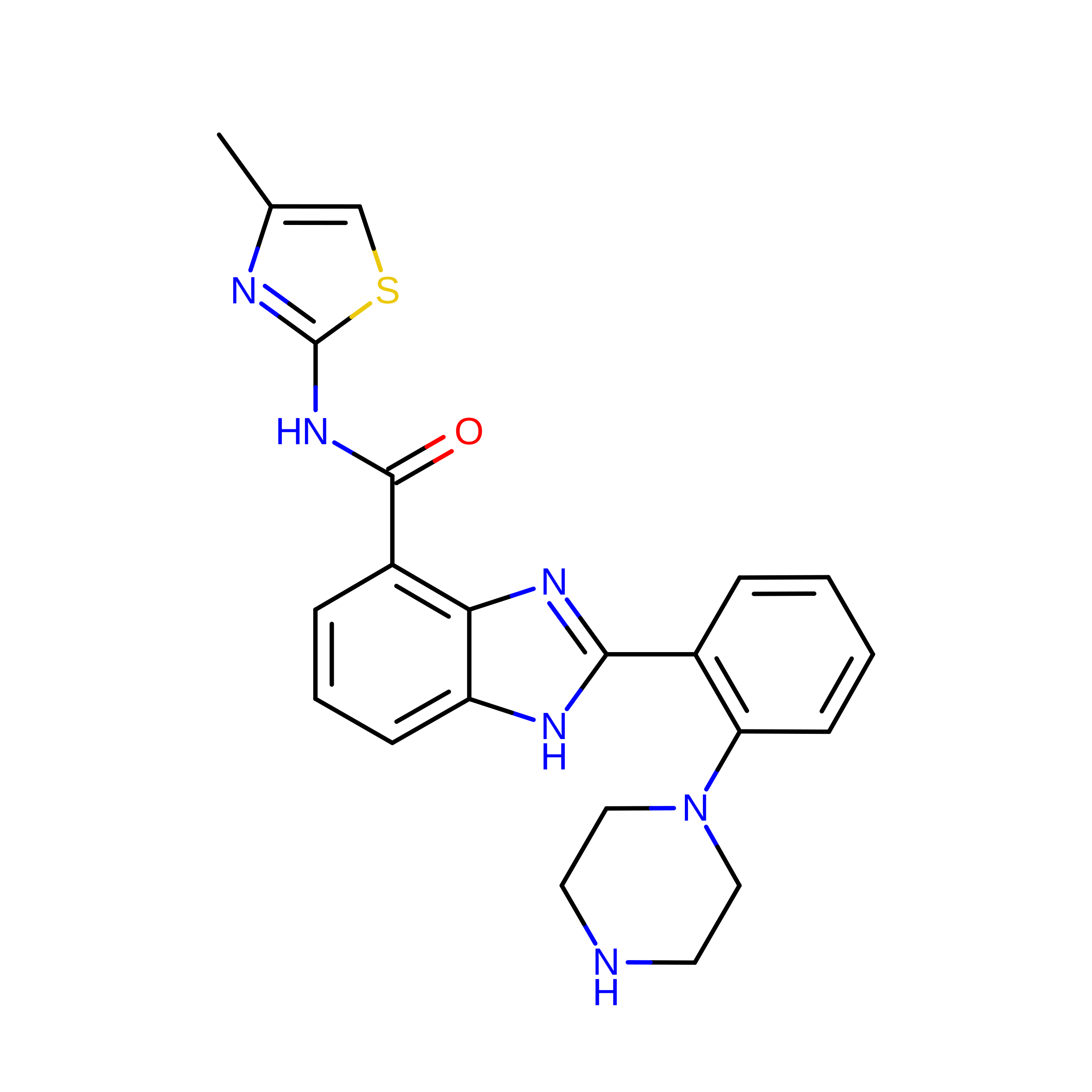 | ||
| 1PE Download:Ideal Coordinates CCD File | H [auth A], I [auth A], P [auth B] | PENTAETHYLENE GLYCOL C10 H22 O6 JLFNLZLINWHATN-UHFFFAOYSA-N | 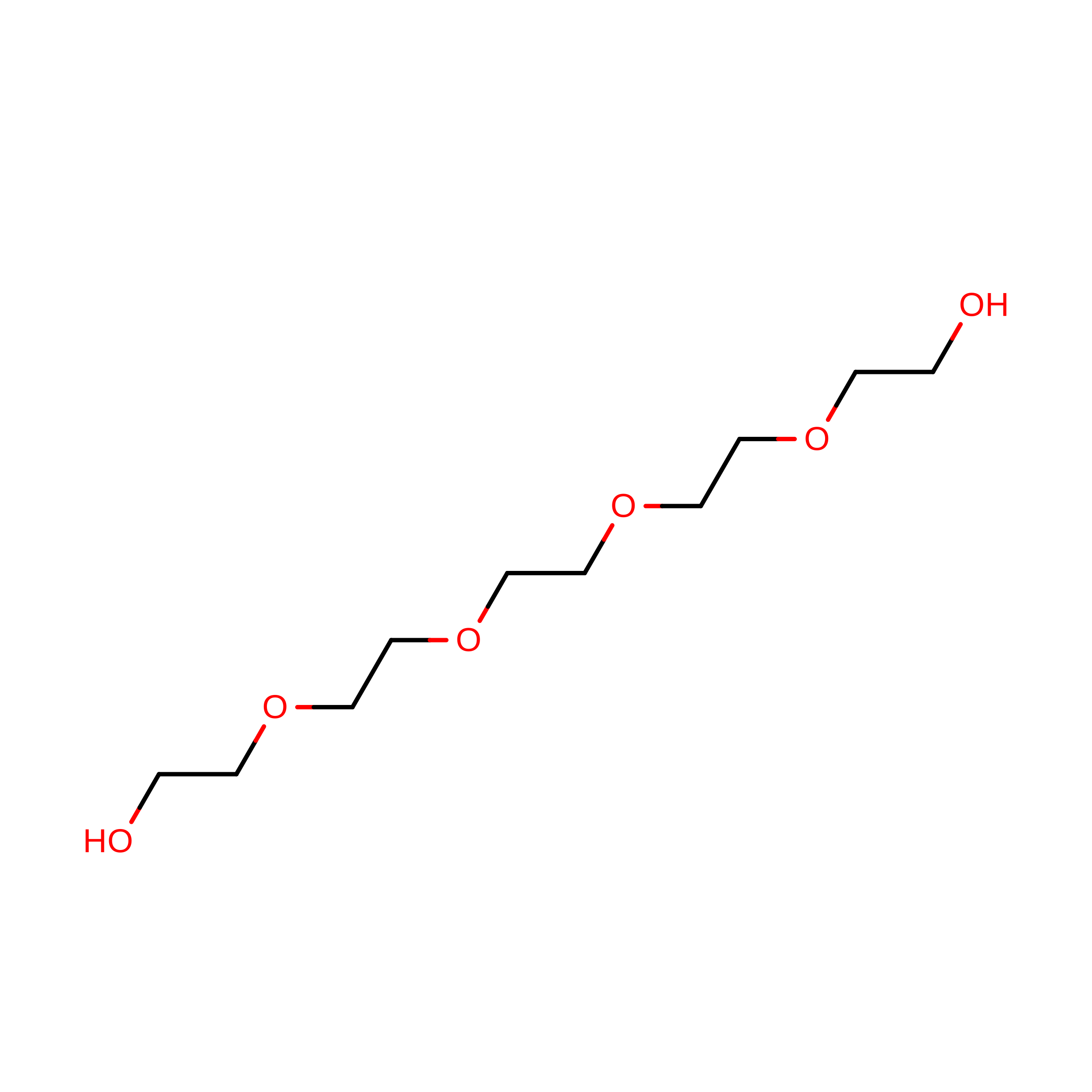 | ||
| PG4 Download:Ideal Coordinates CCD File | G [auth A] | TETRAETHYLENE GLYCOL C8 H18 O5 UWHCKJMYHZGTIT-UHFFFAOYSA-N |  | ||
| PGE Download:Ideal Coordinates CCD File | O [auth B] | TRIETHYLENE GLYCOL C6 H14 O4 ZIBGPFATKBEMQZ-UHFFFAOYSA-N | 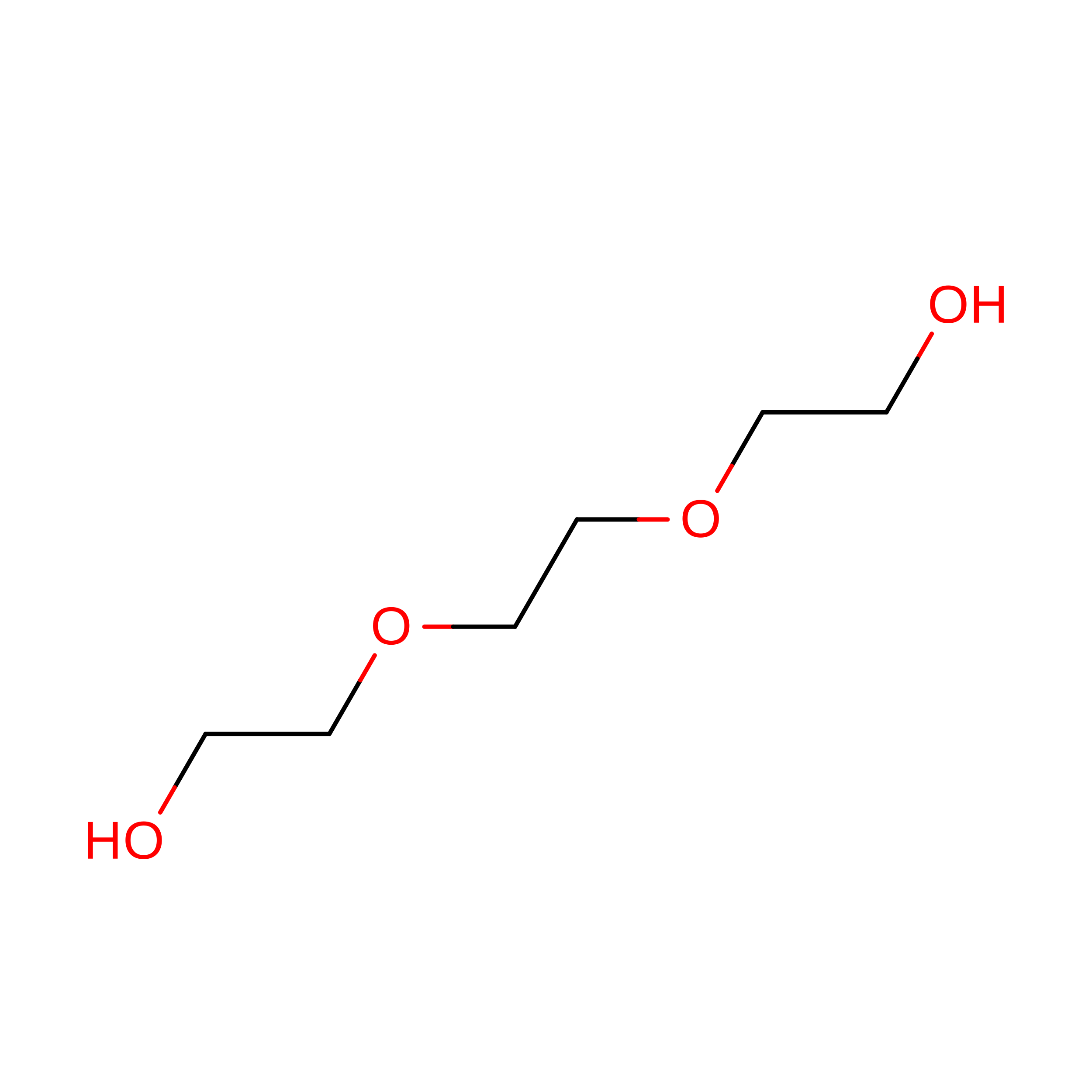 | ||
| CL Download:Ideal Coordinates CCD File | E [auth A] F [auth A] K [auth B] L [auth B] M [auth B] | CHLORIDE ION Cl VEXZGXHMUGYJMC-UHFFFAOYSA-M |  | ||
| Length ( Å ) | Angle ( ˚ ) |
|---|---|
| a = 54.455 | α = 90 |
| b = 88.259 | β = 97.62 |
| c = 98.238 | γ = 90 |
| Software Name | Purpose |
|---|---|
| HKL-2000 | data scaling |
| PHENIX | phasing |
| Coot | model building |
| PHENIX | refinement |
| Funding Organization | Location | Grant Number |
|---|---|---|
| National Institutes of Health/National Institute Of Allergy and Infectious Diseases (NIH/NIAID) | United States | R01 AI155536 |
| National Institutes of Health/National Institute Of Allergy and Infectious Diseases (NIH/NIAID) | United States | 6R21 AI137815-02 |
