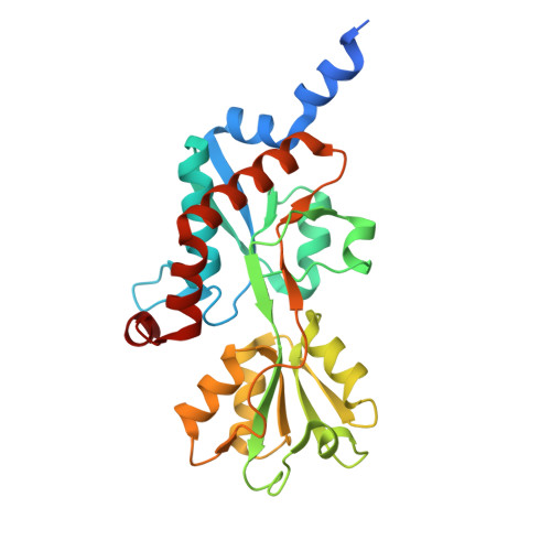
Structure of a solute binding protein from Desulfonauticus sp. bound to L-tryptophan
Rahman, M., Frkic, R.L., Smith, O.B., Jackson, C.J.To be published.
Experimental Data Snapshot
Starting Model: in silico
View more details
| Ligands 5 Unique | |||||
|---|---|---|---|---|---|
| ID | Chains | Name / Formula / InChI Key | 2D Diagram | 3D Interactions | |
| TRP (Subject of Investigation/LOI) Download:Ideal Coordinates CCD File | EA [auth E] G [auth A] KA [auth F] O [auth B] R [auth C] | TRYPTOPHAN C11 H12 N2 O2 QIVBCDIJIAJPQS-VIFPVBQESA-N |  | ||
| PG4 Download:Ideal Coordinates CCD File | FA [auth E] | TETRAETHYLENE GLYCOL C8 H18 O5 UWHCKJMYHZGTIT-UHFFFAOYSA-N |  | ||
| PEG Download:Ideal Coordinates CCD File | Q [auth B], S [auth C], Y [auth D] | DI(HYDROXYETHYL)ETHER C4 H10 O3 MTHSVFCYNBDYFN-UHFFFAOYSA-N |  | ||
| EDO Download:Ideal Coordinates CCD File | AA [auth D] BA [auth D] DA [auth D] GA [auth E] H [auth A] | 1,2-ETHANEDIOL C2 H6 O2 LYCAIKOWRPUZTN-UHFFFAOYSA-N |  | ||
| ACT Download:Ideal Coordinates CCD File | CA [auth D], V [auth C] | ACETATE ION C2 H3 O2 QTBSBXVTEAMEQO-UHFFFAOYSA-M |  | ||
| Length ( Å ) | Angle ( ˚ ) |
|---|---|
| a = 74.507 | α = 98.21 |
| b = 74.593 | β = 91.24 |
| c = 92.348 | γ = 119.84 |
| Software Name | Purpose |
|---|---|
| PHENIX | refinement |
| Aimless | data scaling |
| XDS | data reduction |
| PHASER | phasing |
| Funding Organization | Location | Grant Number |
|---|---|---|
| Australian Research Council (ARC) | Australia | CE200100012 |
| Australian Research Council (ARC) | Australia | CE200100029 |