9DZM | pdb_00009dzm

Dimeric human OCT2 (POU2F2) POU domain bound to palindromic MORE DNA

  • Classification: TRANSCRIPTION/DNA
  • Organism(s): synthetic construct, Homo sapiens
  • Expression System: Escherichia coli
  • Mutation(s): No 

  • Deposited: 2024-10-16 Released: 2024-10-23 
  • Deposition Author(s): Terrell, J.R., Poon, G.M.K.
  • Funding Organization(s): National Institutes of Health/National Heart, Lung, and Blood Institute (NIH/NHLBI), National Science Foundation (NSF, United States), National Institutes of Health/National Institute of General Medical Sciences (NIH/NIGMS)

Experimental Data Snapshot

  • Method: X-RAY DIFFRACTION
  • Resolution: 2.54 Å
  • R-Value Free: 
    0.242 (Depositor), 0.240 (DCC) 
  • R-Value Work: 
    0.217 (Depositor), 0.216 (DCC) 
  • R-Value Observed: 
    0.220 (Depositor) 

Starting Model: experimental
View more details

wwPDB Validation 3D Report Full Report

Validation slider image for 9DZM

This is version 1.2 of the entry. See complete history

Literature

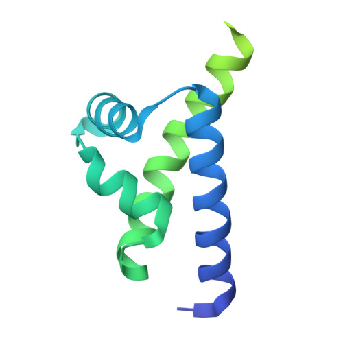
Coupled Heterogeneity to Dimeric Site-Specific Binding by the POU-Family Transcription Factor OCT2.

Terrell, J.R.Poon, G.M.K.

(2025) J Phys Chem B 129: 2138-2148

  • DOI: https://doi.org/10.1021/acs.jpcb.4c07071
  • Primary Citation Related Structures: 
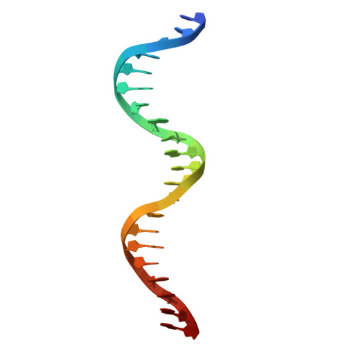
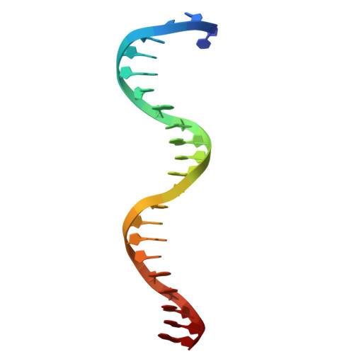
    9DZM

  • PubMed Abstract: 

    POU-family transcription factors regulate metazoan gene expression via a bipartite DNA-binding domain consisting of two covalently linked helix-turn-helix subdomains, POU S and POU H . POU factors bind as dimers to DNA half-sites to form complexes with a variable quaternary structure. To enhance the knowledge of the physical chemistry of dimeric POU/DNA recognition, we carried out a crystallographic and titration analysis of the cooperative homodimer formed by the POU factor OCT2 and an optimized palindromic DNA site known as MORE. The data evidence strong heterogeneity in the binding and formation of secondary complexes in site-specific DNA recognition by OCT2 at thermodynamic equilibrium. These secondary complexes are strictly contingent to the site-specific complex, detectable at subsaturating OCT2 concentrations, and cooperate with nonspecific binding to guide the affinity of the site-specific complex. Modulation with salt and poly[d(I-C)] unmasks the compensation between nonspecific DNA depleting unbound OCT2 on the one hand while driving specific binding by intermolecular transfer of OCT2 via secondary complexes on the other. Molecular dynamics simulations extend a mechanism, previously proposed for POU monomers, in which the two subdomains dynamically cross-link DNA strands to form supramolecular dimeric POU/DNA complexes at equilibrium.


  • Organizational Affiliation
    • Department of Chemistry, Georgia State University, Atlanta, Georgia 30303, United States.

Macromolecule Content 

  • Total Structure Weight: 90.38 kDa 
  • Atom Count: 3,127 
  • Modeled Residue Count: 318 
  • Deposited Residue Count: 711 
  • Unique protein chains: 1
  • Unique nucleic acid chains: 2

Macromolecules


Find similar proteins by:|  3D Structure
Entity ID: 3
MoleculeChains  Sequence LengthOrganismDetailsImage
POU domain, class 2, transcription factor 2
C, D, E, F
167Homo sapiensMutation(s): 0 
Gene Names: POU2F2OCT2OTF2
UniProt & NIH Common Fund Data Resources
Find proteins for P09086 (Homo sapiens)
Explore P09086 
Go to UniProtKB:  P09086
PHAROS:  P09086
GTEx:  ENSG00000028277 
Entity Groups
Sequence Clusters30% Identity50% Identity70% Identity90% Identity95% Identity100% Identity
UniProt GroupP09086
Sequence Annotations
Expand
Reference Sequence
Find similar nucleic acids by:  Sequence
Entity ID: 1
MoleculeChains LengthOrganismImage
DNA (5'-D(*TP*CP*CP*TP*CP*AP*TP*GP*CP*AP*TP*AP*TP*GP*CP*AP*TP*GP*AP*GP*G)-3')21synthetic construct
Sequence Annotations
Expand
Reference Sequence
Find similar nucleic acids by:  Sequence
Entity ID: 2
MoleculeChains LengthOrganismImage
DNA (5'-D(*TP*CP*CP*TP*CP*AP*TP*GP*CP*AP*TP*AP*TP*GP*CP*AP*TP*GP*AP*GP*GP*A)-3')22synthetic construct
Sequence Annotations
Expand
Reference Sequence

Small Molecules

Ligands 1 Unique
IDChains Name / Formula / InChI Key2D Diagram3D Interactions
BR

Query on BR


Download:Ideal Coordinates CCD File
G [auth E]BROMIDE ION
Br
CPELXLSAUQHCOX-UHFFFAOYSA-M

Experimental Data & Validation

Experimental Data

  • Method: X-RAY DIFFRACTION
  • Resolution: 2.54 Å
  • R-Value Free:  0.242 (Depositor), 0.240 (DCC) 
  • R-Value Work:  0.217 (Depositor), 0.216 (DCC) 
  • R-Value Observed: 0.220 (Depositor) 
Space Group: P 1
Unit Cell:
Length ( Å )Angle ( ˚ )
a = 38.029α = 82.373
b = 54.922β = 79.657
c = 69.164γ = 71.769
Software Package:
Software NamePurpose
PHENIXrefinement
XDSdata reduction
Aimlessdata scaling
PHASERphasing

Structure Validation

View Full Validation Report


Entry History 

& Funding Information

Deposition Data


Funding OrganizationLocationGrant Number
National Institutes of Health/National Heart, Lung, and Blood Institute (NIH/NHLBI)United StatesHL155178
National Science Foundation (NSF, United States)United StatesMCB2028902
National Institutes of Health/National Institute of General Medical Sciences (NIH/NIGMS)United StatesGM137160

Revision History  (Full details and data files)

  • Version 1.0: 2024-10-23
    Type: Initial release
  • Version 1.1: 2025-03-05
    Changes: Database references
  • Version 1.2: 2025-03-12
    Changes: Database references