9DUQ | pdb_00009duq

HURP(65-174) bound to GMPCPP-stabilized microtubule


Experimental Data Snapshot

  • Method: ELECTRON MICROSCOPY
  • Resolution: 2.80 Å
  • Aggregation State: FILAMENT 
  • Reconstruction Method: SINGLE PARTICLE 

wwPDB Validation   3D Report Full Report


This is version 1.0 of the entry. See complete history


Literature

HURP facilitates spindle assembly by stabilizing microtubules and working synergistically with TPX2.

Valdez, V.A.Ma, M.Gouveia, B.Zhang, R.Petry, S.

(2024) Nat Commun 15: 9689-9689

  • DOI: https://doi.org/10.1038/s41467-024-53630-6
  • Primary Citation of Related Structures:  
    9DUQ

  • PubMed Abstract: 

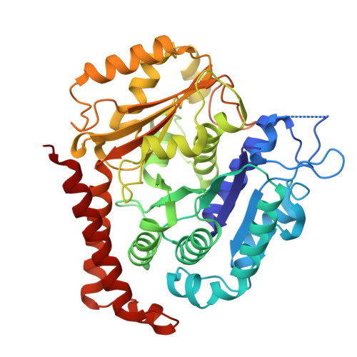
    In vertebrate spindles, most microtubules are formed via branching microtubule nucleation, whereby microtubules nucleate along the side of pre-existing microtubules. Hepatoma up-regulated protein (HURP) is a microtubule-associated protein that has been implicated in spindle assembly, but its mode of action is yet to be defined. In this study, we show that HURP is necessary for RanGTP-induced branching microtubule nucleation in Xenopus egg extract. Specifically, HURP stabilizes the microtubule lattice to promote microtubule formation from γ-TuRC. This function is shifted to promote branching microtubule nucleation through enhanced localization to TPX2 condensates, which form the core of the branch site on microtubules. Lastly, we provide a high-resolution cryo-EM structure of HURP on the microtubule, revealing how HURP binding stabilizes the microtubule lattice. We propose a model in which HURP stabilizes microtubules during their formation, and TPX2 preferentially enriches HURP to microtubules to promote branching microtubule nucleation and thus spindle assembly.


  • Organizational Affiliation
    • Department of Molecular Biology, Princeton University, Princeton, NJ, USA.

Macromolecules
Find similar proteins by:  (by identity cutoff)  |  3D Structure
Entity ID: 1
MoleculeChains Sequence LengthOrganismDetailsImage
Disks large-associated protein 546Homo sapiensMutation(s): 0 
Gene Names: DLGAP5DLG7KIAA0008
UniProt & NIH Common Fund Data Resources
Find proteins for Q15398 (Homo sapiens)
Explore Q15398 
Go to UniProtKB:  Q15398
PHAROS:  Q15398
GTEx:  ENSG00000126787 
Entity Groups  
Sequence Clusters30% Identity50% Identity70% Identity90% Identity95% Identity100% Identity
UniProt GroupQ15398
Sequence Annotations
Expand
  • Reference Sequence
Find similar proteins by:  (by identity cutoff)  |  3D Structure
Entity ID: 2
MoleculeChains Sequence LengthOrganismDetailsImage
Tubulin beta chain
B, D, F, H, J
B, D, F, H, J, L, N, P, R
427Sus scrofaMutation(s): 0 
UniProt
Find proteins for P02554 (Sus scrofa)
Explore P02554 
Go to UniProtKB:  P02554
Entity Groups  
Sequence Clusters30% Identity50% Identity70% Identity90% Identity95% Identity100% Identity
UniProt GroupP02554
Sequence Annotations
Expand
  • Reference Sequence
Find similar proteins by:  (by identity cutoff)  |  3D Structure
Entity ID: 3
MoleculeChains Sequence LengthOrganismDetailsImage
Tubulin alpha chain439Sus scrofaMutation(s): 0 
EC: 3.6.5
UniProt
Find proteins for Q2XVP4 (Sus scrofa)
Explore Q2XVP4 
Go to UniProtKB:  Q2XVP4
Entity Groups  
Sequence Clusters30% Identity50% Identity70% Identity90% Identity95% Identity100% Identity
UniProt GroupQ2XVP4
Sequence Annotations
Expand
  • Reference Sequence
Small Molecules
Ligands 3 Unique
IDChains Name / Formula / InChI Key2D Diagram3D Interactions
GTP
Query on GTP

Download Ideal Coordinates CCD File 
BB [auth M]
DA [auth A]
FB [auth O]
HA [auth C]
JB [auth Q]
BB [auth M],
DA [auth A],
FB [auth O],
HA [auth C],
JB [auth Q],
LA [auth E],
PA [auth G],
TA [auth I],
XA [auth K]
GUANOSINE-5'-TRIPHOSPHATE
C10 H16 N5 O14 P3
XKMLYUALXHKNFT-UUOKFMHZSA-N
G2P
Query on G2P

Download Ideal Coordinates CCD File 
BA [auth B]
DB [auth P]
FA [auth D]
HB [auth R]
JA [auth F]
BA [auth B],
DB [auth P],
FA [auth D],
HB [auth R],
JA [auth F],
NA [auth H],
RA [auth J],
VA [auth L],
ZA [auth N]
PHOSPHOMETHYLPHOSPHONIC ACID GUANYLATE ESTER
C11 H18 N5 O13 P3
GXTIEXDFEKYVGY-KQYNXXCUSA-N
MG
Query on MG

Download Ideal Coordinates CCD File 
AB [auth N]
CA [auth B]
CB [auth M]
EA [auth A]
EB [auth P]
AB [auth N],
CA [auth B],
CB [auth M],
EA [auth A],
EB [auth P],
GA [auth D],
GB [auth O],
IA [auth C],
IB [auth R],
KA [auth F],
KB [auth Q],
MA [auth E],
OA [auth H],
QA [auth G],
SA [auth J],
UA [auth I],
WA [auth L],
YA [auth K]
MAGNESIUM ION
Mg
JLVVSXFLKOJNIY-UHFFFAOYSA-N
Experimental Data & Validation

Experimental Data

  • Method: ELECTRON MICROSCOPY
  • Resolution: 2.80 Å
  • Aggregation State: FILAMENT 
  • Reconstruction Method: SINGLE PARTICLE 
EM Software:
TaskSoftware PackageVersion
RECONSTRUCTIONcryoSPARC3.4
MODEL REFINEMENTPHENIX1.20.1

Structure Validation

View Full Validation Report


Entry History & Funding Information

Deposition Data


Funding OrganizationLocationGrant Number
National Institutes of Health/National Institute of General Medical Sciences (NIH/NIGMS)United StatesR01 GM138854

Revision History  (Full details and data files)

  • Version 1.0: 2024-11-27
    Type: Initial release