Structural insights into SSNA1 self-assembly and its microtubule binding for centriole maintenance.
Agostini, L., Pfister, J.A., Basnet, N., Ding, J., Zhang, R., Biertumpfel, C., O'Connell, K.F., Mizuno, N.(2025) Nat Commun 16: 7512-7512
- PubMed: 40804232 Search on PubMedSearch on PubMed Central
- DOI: https://doi.org/10.1038/s41467-025-62696-9
- Primary Citation Related Structures:
9DSM - PubMed Abstract:
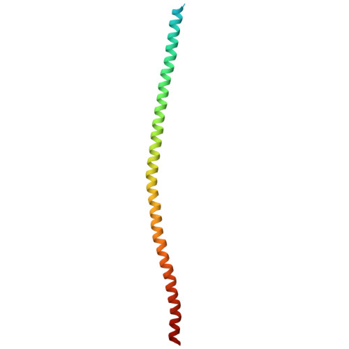
SSNA1 is a fibrillar protein involved in dynamic microtubule remodeling, including nucleation, co-polymerization, and microtubule branching. The underlying molecular mechanism has remained unclear due to a lack of structural information. Here, we determine the cryo-EM structure of C.elegans SSNA-1 at 4.55-Å resolution and evaluate its role in embryonic development. We find that SSNA-1 forms an anti-parallel coiled-coil, with self-assembly facilitated by an overhang of 16 C-terminal residues that form a triple-stranded helical junction. The microtubule-binding region is within the triple-stranded junction, suggesting that self-assembly of SSNA-1 creates hubs for effective microtubule interaction. Genetical analysis elucidates that SSNA-1 deletion significantly reduces embryonic viability, and causes multipolar spindles during cell division. Interestingly, impairing SSNA-1 self-assembly has a comparable effect on embryonic viability as the knockout strain. Our study provides molecular insights into SSNA-1's self-assembly and its role in microtubule binding and cell division regulation through centriole stability.
- Laboratory of Structural Cell Biology, National Heart, Lung, and Blood Institute, National Institutes of Health, 50 South Dr., Bethesda, MD, 20892, USA.
Organizational Affiliation: