


Structural study of sphA
Hai, Y., Xi, W., Chai, W., He, X.To be published.
Experimental Data Snapshot
Starting Model: in silico
View more details
Entity ID: 1 | |||||
|---|---|---|---|---|---|
| Molecule | Chains | Sequence Length | Organism | Details | Image |
| 8-amino-7-oxononanoate synthase | 487 | Aspergillus fumigatus Af293 | Mutation(s): 0 Gene Names: AFUA_3G14690 EC: 2.3.1.47 | 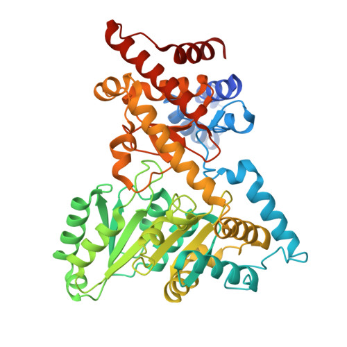 | |
| Ligands 2 Unique | |||||
|---|---|---|---|---|---|
| ID | Chains | Name / Formula / InChI Key | 2D Diagram | 3D Interactions | |
| A1A8V (Subject of Investigation/LOI) Download:Ideal Coordinates CCD File | F [auth A], G [auth B], H [auth C], I [auth D] | (2Z)-2-[({3-hydroxy-2-methyl-5-[(phosphonooxy)methyl]pyridin-4-yl}methyl)imino]butanoic acid C12 H17 N2 O7 P MCYPVVOCEWZTOC-UVTDQMKNSA-N |  | ||
| GOL (Subject of Investigation/LOI) Download:Ideal Coordinates CCD File | E [auth A] | GLYCEROL C3 H8 O3 PEDCQBHIVMGVHV-UHFFFAOYSA-N |  | ||
| Length ( Å ) | Angle ( ˚ ) |
|---|---|
| a = 72.903 | α = 114.902 |
| b = 91.375 | β = 90.144 |
| c = 90.157 | γ = 93.424 |
| Software Name | Purpose |
|---|---|
| PHENIX | refinement |
| HKL-3000 | data reduction |
| HKL-3000 | data scaling |
| PHASER | phasing |
| Funding Organization | Location | Grant Number |
|---|---|---|
| National Institutes of Health/National Institute of General Medical Sciences (NIH/NIGMS) | United States | R35GM151205 |


