9D66 | pdb_00009d66

Human excitatory amino acid transporter 3 (EAAT3) with bound L-Aspartate in an intermediate outward facing state

  • Classification: TRANSPORT PROTEIN
  • Organism(s): Homo sapiens
  • Expression System: Homo sapiens
  • Mutation(s): Yes 
  • Membrane Protein: Yes  PDBTMMemProtMD

  • Deposited: 2024-08-14 Released: 2025-01-22 
  • Deposition Author(s): Qiu, B., Boudker, O.
  • Funding Organization(s): Howard Hughes Medical Institute (HHMI), National Institutes of Health/National Institute of Neurological Disorders and Stroke (NIH/NINDS)

Experimental Data Snapshot

  • Method: ELECTRON MICROSCOPY
  • Resolution: 2.98 Å
  • Aggregation State: PARTICLE 
  • Reconstruction Method: SINGLE PARTICLE 

Starting Model: experimental
View more details

wwPDB Validation   3D Report Full Report


This is version 1.0 of the entry. See complete history


Literature

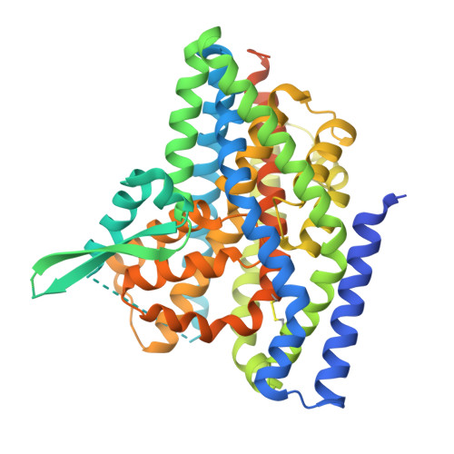
Structural basis of the excitatory amino acid transporter 3 substrate recognition.

Qiu, B.Boudker, O.

(2024) bioRxiv 

  • DOI: https://doi.org/10.1101/2024.09.05.611541
  • Primary Citation of Related Structures:  
    9D66, 9D67, 9D68, 9D69, 9D6A

  • PubMed Abstract: 

    Excitatory amino acid transporters (EAATs) reside on cell surfaces and uptake substrates, including L-glutamate, L-aspartate, and D-aspartate, using ion gradients. Among five EAATs, EAAT3 is the only isoform that can efficiently transport L-cysteine, a substrate for glutathione synthesis. Recent work suggests that EAAT3 also transports the oncometabolite R-2-hydroxyglutarate (R-2HG). Here, we examined the structural basis of substrate promiscuity by determining the cryo-EM structures of EAAT3 bound to different substrates. We found that L-cysteine binds to EAAT3 in thiolate form, and EAAT3 recognizes different substrates by fine-tuning local conformations of the coordinating residues. However, using purified human EAAT3, we could not observe R-2HG binding or transport. Imaging of EAAT3 bound to L-cysteine revealed several conformational states, including an outward-facing state with a semi-open gate and a disrupted sodium-binding site. These structures illustrate that the full gate closure, coupled with the binding of the last sodium ion, occurs after substrate binding. Furthermore, we observed that different substrates affect how the transporter distributes between a fully outward-facing conformation and intermediate occluded states on a path to the inward-facing conformation, suggesting that translocation rates are substrate-dependent.


  • Organizational Affiliation
    • Department of Physiology & Biophysics, Weill Cornell Medicine, 1300 York Ave, New York, NY 10021, USA.

Macromolecules
Find similar proteins by:  (by identity cutoff)  |  3D Structure
Entity ID: 1
MoleculeChains Sequence LengthOrganismDetailsImage
Excitatory amino acid transporter 3
A, B, C
526Homo sapiensMutation(s): 9 
Gene Names: SLC1A1EAAC1EAAT3
Membrane Entity: Yes 
UniProt & NIH Common Fund Data Resources
Find proteins for P43005 (Homo sapiens)
Explore P43005 
Go to UniProtKB:  P43005
PHAROS:  P43005
GTEx:  ENSG00000106688 
Entity Groups  
Sequence Clusters30% Identity50% Identity70% Identity90% Identity95% Identity100% Identity
UniProt GroupP43005
Sequence Annotations
Expand
  • Reference Sequence
Small Molecules
Ligands 3 Unique
IDChains Name / Formula / InChI Key2D Diagram3D Interactions
HG
Query on HG

Download Ideal Coordinates CCD File 
H [auth A],
L [auth B],
R [auth C]
MERCURY (II) ION
Hg
BQPIGGFYSBELGY-UHFFFAOYSA-N
ASP
Query on ASP

Download Ideal Coordinates CCD File 
D [auth A],
K [auth B],
P [auth C]
ASPARTIC ACID
C4 H7 N O4
CKLJMWTZIZZHCS-REOHCLBHSA-N
NA
Query on NA

Download Ideal Coordinates CCD File 
E [auth A]
F [auth A]
G [auth A]
I [auth B]
J [auth B]
E [auth A],
F [auth A],
G [auth A],
I [auth B],
J [auth B],
M [auth B],
N [auth C],
O [auth C],
Q [auth C]
SODIUM ION
Na
FKNQFGJONOIPTF-UHFFFAOYSA-N
Experimental Data & Validation

Experimental Data

  • Method: ELECTRON MICROSCOPY
  • Resolution: 2.98 Å
  • Aggregation State: PARTICLE 
  • Reconstruction Method: SINGLE PARTICLE 
EM Software:
TaskSoftware PackageVersion
MODEL REFINEMENTPHENIX
RECONSTRUCTIONcryoSPARC4

Structure Validation

View Full Validation Report


Entry History & Funding Information

Deposition Data


Funding OrganizationLocationGrant Number
Howard Hughes Medical Institute (HHMI)United States--
National Institutes of Health/National Institute of Neurological Disorders and Stroke (NIH/NINDS)United StatesR37NS085318

Revision History  (Full details and data files)

  • Version 1.0: 2025-01-22
    Type: Initial release