Variations in kinase and effector signaling logic in a bacterial two component signaling network.
Swingle, D., Epstein, L., Aymon, R., Isiorho, E.A., Abzalimov, R.R., Favaro, D.C., Gardner, K.H.(2025) J Biological Chem 301: 108534-108534
- PubMed: 40273983 Search on PubMed
- DOI: https://doi.org/10.1016/j.jbc.2025.108534
- Primary Citation Related Structures:
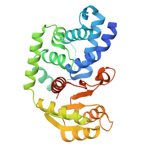
9BY5, 9CB6 - PubMed Abstract:
The general stress response (GSR) protects bacteria from a wide range of stressors. In Alphaproteobacteria, GSR activation is coordinated by HWE/HisKA2 family histidine kinases (HKs), which can exhibit non-canonical structure and function. For example, while most light-oxygen-voltage sensor-containing HKs are light activated dimers, the Rubellimicrobium thermophilum RT-HK has inverted "dark on, light off" signaling logic with a tunable monomer/dimer equilibrium. Here, we further investigate these atypical behaviors of RT-HK and characterize its downstream signaling network. Using hydrogen-deuterium exchange mass spectrometry, we find that RT-HK uses a signal transduction mechanism similar to light-activated systems, despite its inverted logic. Mutagenesis reveals that RT-HK autophosphorylates in trans, with changes to the Jα helix linking sensor and kinase domains affecting autophosphorylation levels. Exploring downstream effects of RT-HK, we identified two GSR genetic regions, each encoding a copy of the central regulator PhyR. In vitro measurements of phosphotransfer from RT-HK to the two putative PhyRs revealed that RT-HK signals only to one, and does so at an increased intensity in the dark, consistent with its reversed logic. X-ray crystal structures of both PhyRs revealed a substantial shift within the receiver domain of one, suggesting a basis for RT-HK specificity. We probed further down the pathway using nuclear magnetic resonance to determine that the single NepR homolog interacts with both unphosphorylated PhyRs, and this interaction is decoupled from activation in one PhyR. This work expands our understanding of HWE/HisKA2 family signal transduction, revealing marked variations from signaling mechanisms previously identified in other GSR networks.
- Structural Biology Initiative, CUNY Advanced Science Research Center, New York, NY 10031; Ph.D. Program in Biochemistry, The Graduate Center - City University of New York, New York, NY 10016.
Organizational Affiliation: