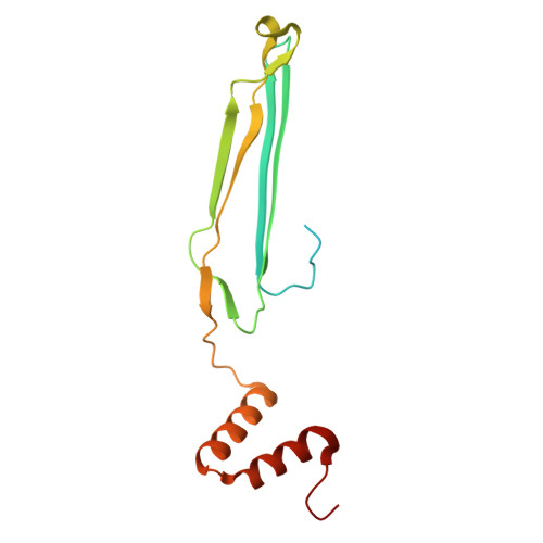
A family of tubular pili from harmful algal bloom forming cyanobacterium Microcystis aeruginosa.
Ricca, J.G., Petersen, H.A., Grosvirt-Dramen, A., Mayali, X., Naylon, S.H., Duersch, B.G., Dufresne, C.P., Weber, P.K., Sonani, R.R., Prevelige, P.E., Hochbaum, A.I., Merk, V., Louda, J.W., Wang, F.(2025) Nat Commun 16: 8082-8082
- PubMed: 40883291 Search on PubMedSearch on PubMed Central
- DOI: https://doi.org/10.1038/s41467-025-63379-1
- Primary Citation Related Structures:
9BTQ - PubMed Abstract:
Cyanobacteria are vital photosynthetic prokaryotes, but some form harmful algal blooms (cyanoHABs) that disrupt ecosystems and produce toxins. The mechanisms by which these blooms form have yet to be fully understood, particularly the role of extracellular components. Here, we present a 2.4 Å cryo-EM structure of a pilus, termed the cyanobacterial tubular (CT) pilus, found in the cyanoHAB-forming Microcystis aeruginosa. The pilin exhibits a unique protein fold, forming a tubular pilus structure with tight, double-layer anti-parallel β-sheet interactions. We show that CT pili are essential for buoyancy by facilitating the formation of micro-colonies, which increases drag force and prevents sinking. The CT pilus surface is heavily glycosylated with ten monosaccharide modifications per pilin. Furthermore, CT pili can enrich microcystin, potentially enhancing cellular resilience, and co-localize with iron-enriched extracellular matrix components. Thus, we propose that this pilus plays an important role in the proliferation of cyanoHABs. This just discovered pilus family appears to be widely distributed across several cyanobacterial orders. Our structural and functional characterization of CT pili provide insights into cyanobacterial cell morphology, physiology, and toxin interactions, and identify potential targets for disrupting bloom formation.
- Department of Chemistry and Biochemistry, Florida Atlantic University, Boca Raton, FL, USA. jgricca@unistra.fr.
Organizational Affiliation: