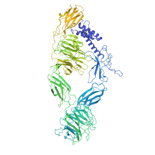
Structural basis for the interaction between the Drosophila RTK Sevenless (dROS1) and the GPCR BOSS.
Zhang, J., Tsutsui, Y., Li, H., Li, T., Wang, Y., Laraki, S., Alarcon-Frias, S., Stayrook, S.E., Klein, D.E.(2025) Nat Commun 16: 808-808
- PubMed: 39827240 Search on PubMedSearch on PubMed Central
- DOI: https://doi.org/10.1038/s41467-025-55943-6
- Primary Citation Related Structures:
9BHX - PubMed Abstract:
Sevenless, the Drosophila homologue of ROS1 (University of Rochester Sarcoma) (herein, dROS1) is a receptor tyrosine kinase (RTK) essential for the differentiation of Drosophila R7 photoreceptor cells. Activation of dROS1 is mediated by binding to the extracellular region (ECR) of the GPCR (G protein coupled receptor) BOSS (Bride Of Sevenless) on adjacent cells. Activation of dROS1 by BOSS leads to subsequent downstream signaling pathways including SOS (Son of Sevenless). However, the physical basis for how dROS1 interacts with BOSS has long remained unknown. Here we provide a cryo-EM structure of dROS1's extracellular region, which mediates ligand binding. We show that the extracellular region of dROS1 adopts a folded-over conformation stabilized by an N-terminal domain comprised of two disulfide stapled helical hairpins. We further narrowed down the interacting binding epitopes on both dROS1 and BOSS using hydrogen-deuterium exchange mass spectrometry (HDX-MS). This includes beta-strands in dROS1's third Fibronectin type III (FNIII) domain and a C-terminal peptide in BOSS' ECR. Our mutagenesis studies, coupled with AlphaFold complex predictions, support a binding interaction mediated by a hydrophobic interaction and beta-strand augmentation between these regions. Our findings provide a fundamental understanding of the regulatory function of dROS1 and further provide mechanistic insight into the human ortholog and oncogene ROS1.
- Department of Pharmacology, Yale University School of Medicine, New Haven, CT, 06520, USA.
Organizational Affiliation: