


Structure of apo Aggregatibacter actinomycetemcomitans SiaP protein
King-Hudson, T.-R.J., Davies, J.S.To be published.
Experimental Data Snapshot
Starting Models: experimental
View more details
Entity ID: 1 | |||||
|---|---|---|---|---|---|
| Molecule | Chains | Sequence Length | Organism | Details | Image |
| DctP family TRAP transporter solute-binding subunit | 306 | Aggregatibacter actinomycetemcomitans | Mutation(s): 0 Gene Names: FXB68_07930 | 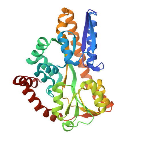 | |
UniProt | |||||
Find proteins for A0A5D0EK58 (Aggregatibacter actinomycetemcomitans) Explore A0A5D0EK58 Go to UniProtKB: A0A5D0EK58 | |||||
Entity Groups | |||||
| Sequence Clusters | 30% Identity50% Identity70% Identity90% Identity95% Identity100% Identity | ||||
| UniProt Group | A0A5D0EK58 | ||||
Sequence AnnotationsExpand | |||||
Reference Sequence | |||||
| Ligands 1 Unique | |||||
|---|---|---|---|---|---|
| ID | Chains | Name / Formula / InChI Key | 2D Diagram | 3D Interactions | |
| ACT (Subject of Investigation/LOI) Download:Ideal Coordinates CCD File | E [auth A], F [auth B] | ACETATE ION C2 H3 O2 QTBSBXVTEAMEQO-UHFFFAOYSA-M |  | ||
| Length ( Å ) | Angle ( ˚ ) |
|---|---|
| a = 138.667 | α = 90 |
| b = 141.394 | β = 112.491 |
| c = 109.405 | γ = 90 |
| Software Name | Purpose |
|---|---|
| PHENIX | refinement |
| XDS | data reduction |
| Aimless | data scaling |
| PHASER | phasing |
| Funding Organization | Location | Grant Number |
|---|---|---|
| Marsden Fund | New Zealand | UOC1506 |


