9BD2 | pdb_00009bd2

MAGE-A3 MHD crystal soaked with KL861


Experimental Data Snapshot

  • Method: X-RAY DIFFRACTION
  • Resolution: 2.24 Å
  • R-Value Free: 
    0.260 (Depositor), 0.258 (DCC) 
  • R-Value Work: 
    0.218 (Depositor), 0.216 (DCC) 
  • R-Value Observed: 
    0.220 (Depositor) 

Starting Model: experimental
View more details

wwPDB Validation   3D Report Full Report


Ligand Structure Quality Assessment 


This is version 1.1 of the entry. See complete history


Literature

Development of Ligands and Degraders Targeting MAGE-A3.

Li, K.Krone, M.W.Butrin, A.Bond, M.J.Linhares, B.M.Crews, C.M.

(2024) J Am Chem Soc 146: 24884-24891

  • DOI: https://doi.org/10.1021/jacs.4c05393
  • Primary Citation of Related Structures:  
    9BD2

  • PubMed Abstract: 

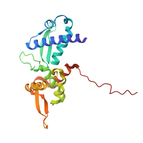
    Type I melanoma antigen (MAGE) family members are detected in numerous tumor types, and expression is correlated with poor prognosis, high tumor grade, and increased metastasis. Type I MAGE proteins are typically restricted to reproductive tissues, but expression can recur during tumorigenesis. Several biochemical functions have been elucidated for them, and notably, MAGEs regulate proteostasis by serving as substrate recognition modules for E3 ligase complexes. The repertoire of E3 ligase complexes that can be hijacked for targeted protein degradation continues to expand, and MAGE-E3 complexes are an especially attractive platform given their cancer-selective expression. Additionally, type I MAGE-derived peptides are presented on cancer cell surfaces, so targeted MAGE degradation may increase antigen presentation and improve immunotherapy outcomes. Motivated by these applications, we developed novel, small-molecule ligands for MAGE-A3, a type I MAGE that is widely expressed in tumors and associates with TRIM28, a RING E3 ligase. Chemical matter was identified through DNA-encoded library (DEL) screening, and hit compounds were validated for in vitro binding to MAGE-A3. We obtained a cocrystal structure with a DEL analog and hypothesize that the small molecule binds at a dimer interface. We utilized this ligand to develop PROTAC molecules that induce MAGE-A3 degradation through VHL recruitment and inhibit the proliferation of MAGE-A3 positive cell lines. These ligands and degraders may serve as valuable probes for investigating MAGE-A3 biology and as foundations for the ongoing development of tumor-specific PROTACs.


  • Organizational Affiliation
    • Department of Molecular, Cellular, and Developmental Biology, Yale University, New Haven, Connecticut 06511, United States.

Macromolecules
Find similar proteins by:  (by identity cutoff)  |  3D Structure
Entity ID: 1
MoleculeChains Sequence LengthOrganismDetailsImage
Melanoma-associated antigen 3
A, B, C
209Homo sapiensMutation(s): 0 
Gene Names: MAGEA3MAGE3
UniProt & NIH Common Fund Data Resources
Find proteins for P43357 (Homo sapiens)
Explore P43357 
Go to UniProtKB:  P43357
PHAROS:  P43357
GTEx:  ENSG00000221867 
Entity Groups  
Sequence Clusters30% Identity50% Identity70% Identity90% Identity95% Identity100% Identity
UniProt GroupP43357
Sequence Annotations
Expand
  • Reference Sequence
Small Molecules
Ligands 1 Unique
IDChains Name / Formula / InChI Key2D Diagram3D Interactions
A1AL3 (Subject of Investigation/LOI)
Query on A1AL3

Download Ideal Coordinates CCD File 
D [auth C](furan-2-yl)[4-({(5P)-5-(1H-indazol-4-yl)-2-[3-(morpholin-4-yl)propoxy]phenyl}methyl)piperazin-1-yl]methanone
C30 H35 N5 O4
SAFWOFNFKMPJAO-UHFFFAOYSA-N
Experimental Data & Validation

Experimental Data

  • Method: X-RAY DIFFRACTION
  • Resolution: 2.24 Å
  • R-Value Free:  0.260 (Depositor), 0.258 (DCC) 
  • R-Value Work:  0.218 (Depositor), 0.216 (DCC) 
  • R-Value Observed: 0.220 (Depositor) 
Space Group: C 2 2 21
Unit Cell:
Length ( Å )Angle ( ˚ )
a = 55.659α = 90
b = 103.402β = 90
c = 325.668γ = 90
Software Package:
Software NamePurpose
PHENIXrefinement
PHENIXrefinement
DIALSdata reduction
Aimlessdata scaling
PHASERphasing

Structure Validation

View Full Validation Report


Ligand Structure Quality Assessment 


Entry History & Funding Information

Deposition Data


Funding OrganizationLocationGrant Number
National Institutes of Health/National Cancer Institute (NIH/NCI)United States--

Revision History  (Full details and data files)

  • Version 1.0: 2024-09-18
    Type: Initial release
  • Version 1.1: 2024-09-25
    Changes: Database references