9BA2 | pdb_00009ba2

Crystal structure of the binary complex of DCAF1 and WDR5


Experimental Data Snapshot

  • Method: X-RAY DIFFRACTION
  • Resolution: 2.97 Å
  • R-Value Free: 
    0.266 (Depositor), 0.219 (DCC) 
  • R-Value Work: 
    0.230 (Depositor) 
  • R-Value Observed: 
    0.231 (Depositor) 

Starting Models: experimental
View more details

wwPDB Validation 3D Report Full Report

Validation slider image for 9BA2

Ligand Structure Quality Assessment 


This is version 1.1 of the entry. See complete history

Literature

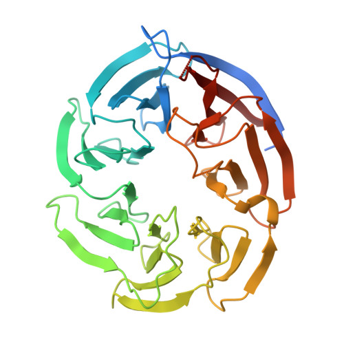
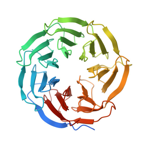
Crystal structures of DCAF1-PROTAC-WDR5 ternary complexes provide insight into DCAF1 substrate specificity.

Mabanglo, M.F.Wilson, B.Noureldin, M.Kimani, S.W.Mamai, A.Krausser, C.Gonzalez-Alvarez, H.Srivastava, S.Mohammed, M.Hoffer, L.Chan, M.Avrumutsoae, J.Li, A.S.M.Hajian, T.Tucker, S.Green, S.Szewczyk, M.Barsyte-Lovejoy, D.Santhakumar, V.Ackloo, S.Loppnau, P.Li, Y.Seitova, A.Kiyota, T.Wang, J.G.Prive, G.G.Kuntz, D.A.Patel, B.Rathod, V.Vala, A.Rout, B.Aman, A.Poda, G.Uehling, D.Ramnauth, J.Halabelian, L.Marcellus, R.Al-Awar, R.Vedadi, M.

(2024) Nat Commun 15: 10165-10165

  • DOI: https://doi.org/10.1038/s41467-024-54500-x
  • Primary Citation Related Structures: 
    9B9H, 9B9T, 9B9W, 9BA2, 9DLW

  • PubMed Abstract: 

    Proteolysis-targeting chimeras (PROTACs) have been explored for the degradation of drug targets for more than two decades. However, only a handful of E3 ligase substrate receptors have been efficiently used. Downregulation and mutation of these receptors would reduce the effectiveness of such PROTACs. We recently developed potent ligands for DCAF1, a substrate receptor of EDVP and CUL4 E3 ligases. Here, we focus on DCAF1 toward the development of PROTACs for WDR5, a drug target in various cancers. We report four DCAF1-based PROTACs with endogenous and exogenous WDR5 degradation effects and high-resolution crystal structures of the ternary complexes of DCAF1-PROTAC-WDR5. The structures reveal detailed insights into the interaction of DCAF1 with various WDR5-PROTACs, indicating a significant role of DCAF1 loops in providing needed surface plasticity, and reflecting the mechanism by which DCAF1 functions as a substrate receptor for E3 ligases with diverse sets of substrates.


  • Organizational Affiliation
    • Drug Discovery Program, Ontario Institute for Cancer Research, Toronto, ON, Canada.

Macromolecule Content 

  • Total Structure Weight: 74.8 kDa 
  • Atom Count: 4,887 
  • Modeled Residue Count: 611 
  • Deposited Residue Count: 667 
  • Unique protein chains: 2

Macromolecules

Find similar proteins by:|  3D Structure
Entity ID: 1
MoleculeChains  Sequence LengthOrganismDetailsImage
DDB1- and CUL4-associated factor 1338Homo sapiensMutation(s): 0 
Gene Names: DCAF1KIAA0800RIPVPRBP
EC: 2.7.11.1
UniProt & NIH Common Fund Data Resources
Find proteins for Q9Y4B6 (Homo sapiens)
Explore Q9Y4B6 
Go to UniProtKB:  Q9Y4B6
PHAROS:  Q9Y4B6
GTEx:  ENSG00000145041 
Entity Groups
Sequence Clusters30% Identity50% Identity70% Identity90% Identity95% Identity100% Identity
UniProt GroupQ9Y4B6
Sequence Annotations
Expand
Reference Sequence
Find similar proteins by:|  3D Structure
Entity ID: 2
MoleculeChains  Sequence LengthOrganismDetailsImage
WD repeat-containing protein 5329Homo sapiensMutation(s): 0 
Gene Names: WDR5BIG3
UniProt & NIH Common Fund Data Resources
Find proteins for P61964 (Homo sapiens)
Explore P61964 
Go to UniProtKB:  P61964
PHAROS:  P61964
GTEx:  ENSG00000196363 
Entity Groups
Sequence Clusters30% Identity50% Identity70% Identity90% Identity95% Identity100% Identity
UniProt GroupP61964
Sequence Annotations
Expand
Reference Sequence

Small Molecules

Ligands 1 Unique
IDChains Name / Formula / InChI Key2D Diagram3D Interactions
IMD
(Subject of Investigation/LOI)

Query on IMD


Download:Ideal Coordinates CCD File
C [auth A]IMIDAZOLE
C3 H5 N2
RAXXELZNTBOGNW-UHFFFAOYSA-O

Experimental Data & Validation

Experimental Data

  • Method: X-RAY DIFFRACTION
  • Resolution: 2.97 Å
  • R-Value Free:  0.266 (Depositor), 0.219 (DCC) 
  • R-Value Work:  0.230 (Depositor) 
  • R-Value Observed: 0.231 (Depositor) 
Space Group: P 21 21 21
Unit Cell:
Length ( Å )Angle ( ˚ )
a = 47.597α = 90
b = 83.185β = 90
c = 158.152γ = 90
Software Package:
Software NamePurpose
PHENIXrefinement
XDSdata reduction
pointlessdata scaling
PHASERphasing

Structure Validation

View Full Validation Report


Ligand Structure Quality Assessment 


Entry History 

& Funding Information

Deposition Data


Funding OrganizationLocationGrant Number
Ontario Institute for Cancer ResearchCanada--

Revision History  (Full details and data files)

  • Version 1.0: 2024-11-06
    Type: Initial release
  • Version 1.1: 2024-12-04
    Changes: Database references