8ZUE | pdb_00008zue

cryo-electron microscopy (cryo-EM) structure of the Hachiman defense system from Escherichia coli


Experimental Data Snapshot

  • Method: ELECTRON MICROSCOPY
  • Resolution: 3.10 Å
  • Aggregation State: PARTICLE 
  • Reconstruction Method: SINGLE PARTICLE 

wwPDB Validation   3D Report Full Report


This is version 1.1 of the entry. See complete history


Literature

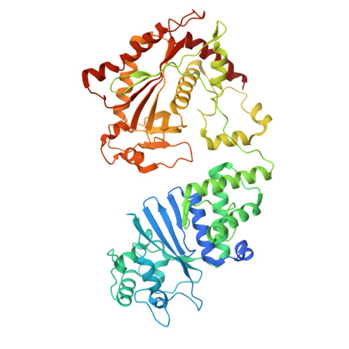
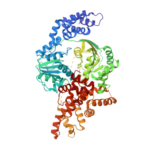
Bacterial Hachiman complex executes DNA cleavage for antiphage defense.

Cui, Y.Dai, Z.Ouyang, Y.Fu, C.Wang, Y.Chen, X.Yang, K.Zheng, S.Wang, W.Tao, P.Guan, Z.Zou, T.

(2025) Nat Commun 16: 2604-2604

  • DOI: https://doi.org/10.1038/s41467-025-57851-1
  • Primary Citation of Related Structures:  
    8ZUE

  • PubMed Abstract: 

    Bacteria have developed a variety of immune systems to combat phage infections. The Hachiman system is a novel prokaryotic antiphage defense system comprising HamA and HamB proteins, which contains the DUF1837 and helicase domains, respectively. However, the defense mechanism remains only partially understood. Here, we present the cryo-electron microscopy (cryo-EM) structure of the Hachiman defense system featuring a fusion of Cap4 nuclease domain within HamA. Further structure analysis indicates that the DUF1837 domain on HamA resembles the PD-(D/E)XK nuclease but lacks active sites. Bioinformatics analysis reveals that catalytically inactive DUF1837 domains often recruit other functional domains to fulfill anti-phage defense. HamA interacts with HamB to form a heterodimer HamAB to mediate ATP hydrolysis and execute DNA cleavage, thus implementing antiphage defense. Our findings elucidate the structural basis of the Hachiman defense complex, highlighting the critical roles of the helicase and nuclease in prokaryotic immunity.


  • Organizational Affiliation
    • National Key Laboratory of Agricultural Microbiology, Hubei Hongshan Laboratory, Huazhong Agricultural University, Wuhan, China.

Macromolecules
Find similar proteins by:  (by identity cutoff)  |  3D Structure
Entity ID: 1
MoleculeChains Sequence LengthOrganismDetailsImage
Anti-bacteriophage protein A565Escherichia coli K-12Mutation(s): 0 
Gene Names: abpAyfjLb2628JW2609
UniProt
Find proteins for P52127 (Escherichia coli (strain K12))
Explore P52127 
Go to UniProtKB:  P52127
Entity Groups  
Sequence Clusters30% Identity50% Identity70% Identity90% Identity95% Identity100% Identity
UniProt GroupP52127
Sequence Annotations
Expand
  • Reference Sequence
Find similar proteins by:  (by identity cutoff)  |  3D Structure
Entity ID: 2
MoleculeChains Sequence LengthOrganismDetailsImage
Anti-bacteriophage protein B729Escherichia coli K-12Mutation(s): 0 
Gene Names: abpByfjKb2627JW2608
UniProt
Find proteins for P52126 (Escherichia coli (strain K12))
Explore P52126 
Go to UniProtKB:  P52126
Entity Groups  
Sequence Clusters30% Identity50% Identity70% Identity90% Identity95% Identity100% Identity
UniProt GroupP52126
Sequence Annotations
Expand
  • Reference Sequence
Experimental Data & Validation

Experimental Data

  • Method: ELECTRON MICROSCOPY
  • Resolution: 3.10 Å
  • Aggregation State: PARTICLE 
  • Reconstruction Method: SINGLE PARTICLE 
EM Software:
TaskSoftware PackageVersion
MODEL REFINEMENTPHENIX

Structure Validation

View Full Validation Report


Entry History & Funding Information

Deposition Data


Funding OrganizationLocationGrant Number
National Natural Science Foundation of China (NSFC)China--

Revision History  (Full details and data files)

  • Version 1.0: 2025-03-26
    Type: Initial release
  • Version 1.1: 2025-07-23
    Changes: Data collection