8ZQG | pdb_00008zqg

Crystal structure of WIPI3 in complex with ATG16L1


Experimental Data Snapshot

  • Method: X-RAY DIFFRACTION
  • Resolution: 2.77 Å
  • R-Value Free: 
    0.233 (Depositor), 0.231 (DCC) 
  • R-Value Work: 
    0.190 (Depositor), 0.189 (DCC) 
  • R-Value Observed: 
    0.192 (Depositor) 

Starting Model: experimental
View more details

wwPDB Validation   3D Report Full Report


This is version 1.1 of the entry. See complete history


Literature

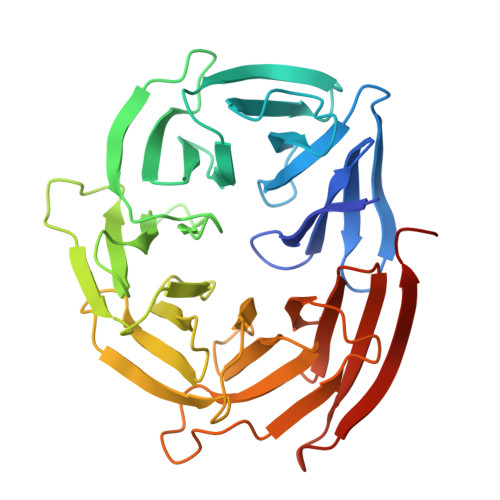
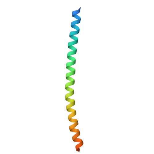
Structure of the WIPI3/ATG16L1 Complex Reveals the Molecular Basis for the Recruitment of the ATG12~ATG5-ATG16L1 Complex by WIPI3.

Gong, X.Wang, Y.Zhou, Y.Pan, L.

(2024) Cells 13

  • DOI: https://doi.org/10.3390/cells13242113
  • Primary Citation of Related Structures:  
    8ZQG

  • PubMed Abstract: 

    Macroautophagy deploys a wealth of autophagy-related proteins to synthesize the double-membrane autophagosome, in order to engulf cytosolic components for lysosome-dependent degradation. The recruitment of the ATG12~ATG5-ATG16L1 complex by WIPI family proteins is a crucial step in autophagosome formation. Nevertheless, the molecular mechanism by which WIPI3 facilitates the recruitment of the ATG12~ATG5-ATG16L1 complex remains largely unknown. Here, we uncover that WIPI3 can directly interact with the coiled-coil domain of ATG16L1. By determining the crystal structure of WIPI3 in complex with ATG16L1 coiled-coil, we elucidate the molecular basis underpinning the specific recruitment of the ATG12~ATG5-ATG16L1 complex by WIPI3. Moreover, we demonstrate that WIPI2 and WIPI3 are competitive for interacting with ATG16L1 coiled-coil, and ATG16L1 and ATG2 are mutually exclusive in binding to WIPI3. In all, our findings provide mechanistic insights into the WIPI3/ATG16L1 interaction, and are valuable for further understanding the activation mechanism of the ATG12~ATG5-ATG16L1 complex as well as the working mode of WIPI3 in autophagy.


  • Organizational Affiliation
    • State Key Laboratory of Chemical Biology, Shanghai Institute of Organic Chemistry, University of Chinese Academy of Sciences, Chinese Academy of Sciences, Shanghai 200032, China.

Macromolecules
Find similar proteins by:  (by identity cutoff)  |  3D Structure
Entity ID: 1
MoleculeChains Sequence LengthOrganismDetailsImage
WD repeat domain phosphoinositide-interacting protein 3A [auth B],
D [auth A]
323Homo sapiensMutation(s): 0 
Gene Names: WDR45BWDR45LWIPI3
UniProt & NIH Common Fund Data Resources
Find proteins for Q5MNZ6 (Homo sapiens)
Explore Q5MNZ6 
Go to UniProtKB:  Q5MNZ6
PHAROS:  Q5MNZ6
GTEx:  ENSG00000141580 
Entity Groups  
Sequence Clusters30% Identity50% Identity70% Identity90% Identity95% Identity100% Identity
UniProt GroupQ5MNZ6
Sequence Annotations
Expand
  • Reference Sequence
Find similar proteins by:  (by identity cutoff)  |  3D Structure
Entity ID: 2
MoleculeChains Sequence LengthOrganismDetailsImage
Autophagy-related protein 16-1B [auth C],
C [auth D]
69Homo sapiensMutation(s): 0 
Gene Names: ATG16L1APG16LUNQ9393/PRO34307
UniProt & NIH Common Fund Data Resources
Find proteins for Q676U5 (Homo sapiens)
Explore Q676U5 
Go to UniProtKB:  Q676U5
PHAROS:  Q676U5
GTEx:  ENSG00000085978 
Entity Groups  
Sequence Clusters30% Identity50% Identity70% Identity90% Identity95% Identity100% Identity
UniProt GroupQ676U5
Sequence Annotations
Expand
  • Reference Sequence
Experimental Data & Validation

Experimental Data

  • Method: X-RAY DIFFRACTION
  • Resolution: 2.77 Å
  • R-Value Free:  0.233 (Depositor), 0.231 (DCC) 
  • R-Value Work:  0.190 (Depositor), 0.189 (DCC) 
  • R-Value Observed: 0.192 (Depositor) 
Space Group: P 63
Unit Cell:
Length ( Å )Angle ( ˚ )
a = 182.663α = 90
b = 182.663β = 90
c = 54.34γ = 120
Software Package:
Software NamePurpose
PHENIXrefinement
autoPROCdata reduction
autoPROCdata scaling
PHENIXphasing

Structure Validation

View Full Validation Report


Entry History & Funding Information

Deposition Data


Funding OrganizationLocationGrant Number
National Natural Science Foundation of China (NSFC)China21822705

Revision History  (Full details and data files)

  • Version 1.0: 2025-01-01
    Type: Initial release
  • Version 1.1: 2025-01-22
    Changes: Database references