


Crystal structure of a novel alginate-binding carbohydrate binding module
Mei, X.W., Tao, W.W., Chang, Y.G.To be published.
Experimental Data Snapshot
Starting Model: in silico
View more details
wwPDB Validation 3D Report Full Report

Entity ID: 1 | |||||
|---|---|---|---|---|---|
| Molecule | Chains | Sequence Length | Organism | Details | Image |
| Alginate lyase | 214 | Vibrio breoganii | Mutation(s): 0 Gene Names: A6E01_07505 | 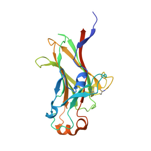 | |
| Length ( Å ) | Angle ( ˚ ) |
|---|---|
| a = 38.95 | α = 90 |
| b = 64.36 | β = 110.8 |
| c = 41.44 | γ = 90 |
| Software Name | Purpose |
|---|---|
| PHENIX | refinement |
| Aimless | data scaling |
| XDS | data reduction |
| PHASER | phasing |
| PDB_EXTRACT | data extraction |
| Funding Organization | Location | Grant Number |
|---|---|---|
| Not funded | -- |


