8ZMD | pdb_00008zmd

Protease-activated receptor-2 (PAR2)/Gq complex


Experimental Data Snapshot

  • Method: ELECTRON MICROSCOPY
  • Resolution: 3.25 Å
  • Aggregation State: PARTICLE 
  • Reconstruction Method: SINGLE PARTICLE 

wwPDB Validation   3D Report Full Report


This is version 1.2 of the entry. See complete history


Literature

Structural basis of protease-activated receptor 2 activation and biased agonism.

Zhu, X.Xia, R.Zhang, A.Guo, C.Xu, Z.He, Y.

(2025) Cell Discov 11: 96-96

  • DOI: https://doi.org/10.1038/s41421-025-00851-8
  • Primary Citation of Related Structures:  
    8ZMD, 8ZME

  • PubMed Abstract: 

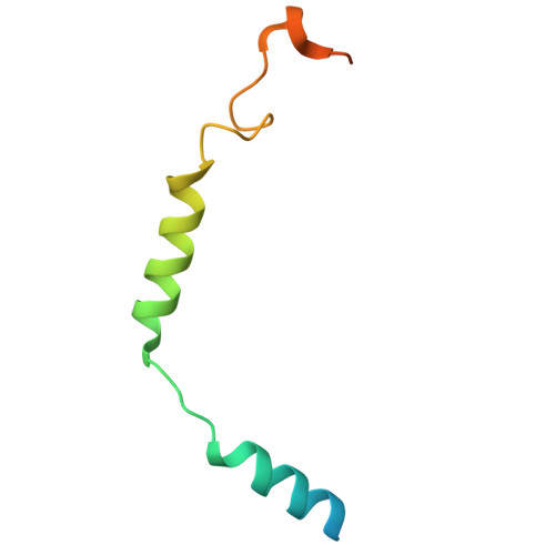
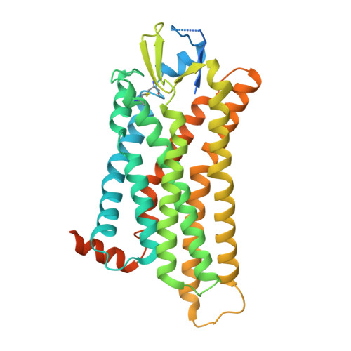
    Protease-activated receptor 2 (PAR2) is a transmembrane receptor that is irreversibly activated by proteolytic cleavage of its N-terminus via extracellular proteases, resulting in the release of the tethered ligand (TL), which binds to and activates the receptor. PAR2 plays a pivotal role in the inflammatory response and pain sensation and is a promising drug target for treating arthritis, asthma, and neuronal pain. Here, we present the cryo-electron microscopy structures of active PAR2 complexed with miniG s/q and miniG 13 . Combining functional assays with structural analysis, our study revealed that TL forms a parallel β-sheet with the extracellular loop 2 of PAR2 to engage the receptor. The binding of TL triggers a conformational rearrangement in the transmembrane core, releasing the inhibitory ion lock and allowing receptor activation. Furthermore, we provide structural insights into the engagement of G q and G 13 with PAR2, highlighting that a hydrophobic interaction mediated by the last methionine residue of Gα 13 is crucial for G 13 coupling selectivity. In combination with molecular dynamics simulations and mutagenesis, we identified the I39 TL3 /D62 N-term interaction at the pocket side of the receptor as a key determinant of G 13 signaling. Disrupting this interaction significantly inhibits G 13 signaling while preserving G q activity, enabling us to design a biased peptide ligand that selectively activates G q signaling. The information revealed in this study provides a framework for understanding PAR2 signaling and offers a rational basis for the design of biased PAR2 ligands.


  • Organizational Affiliation
    • HIT Center for Life Sciences, School of Life Science and Technology, Faculty of Life Sciences and Medicine, Harbin Institute of Technology, Harbin, Heilongjiang, China.

Macromolecules
Find similar proteins by:  (by identity cutoff)  |  3D Structure
Entity ID: 1
MoleculeChains Sequence LengthOrganismDetailsImage
engineered G alpha q362Homo sapiensMutation(s): 0 
Entity Groups  
Sequence Clusters30% Identity50% Identity70% Identity90% Identity95% Identity100% Identity
Sequence Annotations
Expand
  • Reference Sequence
Find similar proteins by:  (by identity cutoff)  |  3D Structure
Entity ID: 2
MoleculeChains Sequence LengthOrganismDetailsImage
Guanine nucleotide-binding protein G(I)/G(S)/G(T) subunit beta-1345Homo sapiensMutation(s): 0 
Gene Names: GNB1
UniProt & NIH Common Fund Data Resources
Find proteins for P62873 (Homo sapiens)
Explore P62873 
Go to UniProtKB:  P62873
PHAROS:  P62873
GTEx:  ENSG00000078369 
Entity Groups  
Sequence Clusters30% Identity50% Identity70% Identity90% Identity95% Identity100% Identity
UniProt GroupP62873
Sequence Annotations
Expand
  • Reference Sequence
Find similar proteins by:  (by identity cutoff)  |  3D Structure
Entity ID: 3
MoleculeChains Sequence LengthOrganismDetailsImage
Guanine nucleotide-binding protein G(I)/G(S)/G(O) subunit gamma-2C [auth G]71Homo sapiensMutation(s): 0 
Gene Names: GNG2
Membrane Entity: Yes 
UniProt & NIH Common Fund Data Resources
Find proteins for P59768 (Homo sapiens)
Explore P59768 
Go to UniProtKB:  P59768
PHAROS:  P59768
GTEx:  ENSG00000186469 
Entity Groups  
Sequence Clusters30% Identity50% Identity70% Identity90% Identity95% Identity100% Identity
UniProt GroupP59768
Sequence Annotations
Expand
  • Reference Sequence
Find similar proteins by:  (by identity cutoff)  |  3D Structure
Entity ID: 4
MoleculeChains Sequence LengthOrganismDetailsImage
Proteinase-activated receptor 2D [auth R]397Homo sapiensMutation(s): 0 
Gene Names: F2RL1GPR11PAR2
Membrane Entity: Yes 
UniProt & NIH Common Fund Data Resources
Find proteins for P55085 (Homo sapiens)
Explore P55085 
Go to UniProtKB:  P55085
PHAROS:  P55085
GTEx:  ENSG00000164251 
Entity Groups  
Sequence Clusters30% Identity50% Identity70% Identity90% Identity95% Identity100% Identity
UniProt GroupP55085
Sequence Annotations
Expand
  • Reference Sequence
Experimental Data & Validation

Experimental Data

  • Method: ELECTRON MICROSCOPY
  • Resolution: 3.25 Å
  • Aggregation State: PARTICLE 
  • Reconstruction Method: SINGLE PARTICLE 
EM Software:
TaskSoftware PackageVersion
MODEL REFINEMENTPHENIX

Structure Validation

View Full Validation Report


Entry History & Funding Information

Deposition Data

  • Released Date: 2025-05-28 
  • Deposition Author(s): He, Y., Zhu, X.

Funding OrganizationLocationGrant Number
National Natural Science Foundation of China (NSFC)China32070048

Revision History  (Full details and data files)

  • Version 1.0: 2025-05-28
    Type: Initial release
  • Version 1.1: 2025-07-23
    Changes: Data collection
  • Version 1.2: 2025-12-10
    Changes: Data collection, Database references