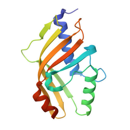
8ZED | pdb_00008zed

Crystal structure of aldolase AtoB


Experimental Data Snapshot

  • Method: X-RAY DIFFRACTION
  • Resolution: 2.37 Å
  • R-Value Free: 
    0.321 (Depositor), 0.321 (DCC) 
  • R-Value Work: 
    0.260 (Depositor), 0.260 (DCC) 
  • R-Value Observed: 
    0.263 (Depositor) 

Starting Model: experimental
View more details

wwPDB Validation 3D Report Full Report

Validation slider image for 8ZED

This is version 1.1 of the entry. See complete history. 

Literature

Three-dimensional structural alignment based discovery and molecular basis of AtoB, catalyzing linear tetracyclic formation.

Ma, K., Liu, J., Huang, Z., Wu, M., Liu, D., Ren, J., Fan, A., Lin, W.

(2024) Chem Sci 15: 18490-18496

  • DOI: https://doi.org/10.1039/d4sc05590j
  • Primary Citation Related Structures: 
    8ZEC, 8ZED, 9JLM

  • PubMed Abstract: 

    Enzymes from the nuclear transport factor 2-like (NTF2-like) superfamily represent a rare group of biocatalysts with diverse catalytic functions facilitating intriguing skeleton formations. However, most proteins of this family remain enigmatic and await further elucidation. In this study, a combination of protein structural alignment with clustering analysis uncovers a new aldolase, AtoB, belonging to the NTF2-like superfamily. AtoB catalyzes the key intramolecular aldol reaction in linear tetracyclic meroterpenoid biosynthesis. The X-ray crystal structures of AtoB and AtoB-ligand complex are established at 1.9 Å and 1.6 Å resolution, respectively, revealing the rotation of the α4 helix and key residues in the active site for substrate binding. Molecular docking and site-directed mutagenesis demonstrate an acid-base pair involved in the AtoB-catalyzed aldol reaction, of which Arg59 is responsible for stereocontrol of hydroxylated C-10a during condensation. These findings provide valuable information for understanding the catalytic mechanisms of the AtoB-catalyzed aldol reaction. Additionally, a branching biosynthetic pathway of aspertetranones is elucidated during the exploration of the natural substrate of AtoB.


  • Organizational Affiliation: 
    • State Key Laboratory of Natural and Biomimetic Drugs, School of Pharmaceutical Sciences, Peking University Beijing 100191 China fanaili@bjmu.edu.cn whlin@bjmu.edu.cn.

Macromolecule Content 

  • Total Structure Weight: 66.82 kDa 
  • Atom Count: 4,585 
  • Modeled Residue Count: 552 
  • Deposited Residue Count: 588 
  • Unique protein chains: 1

Macromolecules

Find similar proteins by:|  3D Structure
Entity ID: 1
MoleculeChains  Sequence LengthOrganismDetailsImage
AtoB aldolase
A, B, C, D
147Aspergillus ochraceusMutation(s): 0 
Gene Names: atoB
UniProt
Find proteins for A0A0U5GHC1 (Aspergillus calidoustus)
Explore A0A0U5GHC1 
Go to UniProtKB:  A0A0U5GHC1
Entity Groups
Sequence Clusters30% Identity50% Identity70% Identity90% Identity95% Identity100% Identity
UniProt GroupA0A0U5GHC1
Sequence Annotations
Expand
Reference Sequence

Experimental Data & Validation

Experimental Data

  • Method: X-RAY DIFFRACTION
  • Resolution: 2.37 Å
  • R-Value Free:  0.321 (Depositor), 0.321 (DCC) 
  • R-Value Work:  0.260 (Depositor), 0.260 (DCC) 
  • R-Value Observed: 0.263 (Depositor) 
Space Group: P 1 21 1
Unit Cell:
Length ( Å )Angle ( ˚ )
a = 51.562α = 90
b = 122.893β = 90
c = 69.728γ = 90
Software Package:
Software NamePurpose
PHENIXrefinement
PHENIXrefinement
HKL-3000data reduction
HKL-3000data scaling
PHASERphasing

Structure Validation

View Full Validation Report


Entry History 

& Funding Information

Deposition Data


Funding OrganizationLocationGrant Number
National Natural Science Foundation of China (NSFC)China82104056

Revision History  (Full details and data files)

  • Version 1.0: 2025-05-14
    Type: Initial release
  • Version 1.1: 2025-11-26
    Changes: Database references