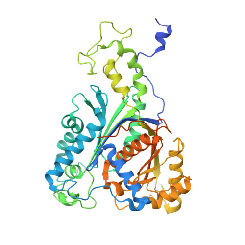
8YZ3 | pdb_00008yz3

Crystal structure of Type Six Secretion System adaptor protein Tla3 from Pseudomonas aeruginosa.


Experimental Data Snapshot

  • Method: X-RAY DIFFRACTION
  • Resolution: 1.80 Å
  • R-Value Free: 
    0.203 (Depositor), 0.204 (DCC) 
  • R-Value Work: 
    0.175 (Depositor), 0.176 (DCC) 
  • R-Value Observed: 
    0.176 (Depositor) 

wwPDB Validation 3D Report Full Report

Validation slider image for 8YZ3

This is version 1.3 of the entry. See complete history

Literature

Crystal structure of the type VI secretion system adaptor protein Tla3 from Pseudomonas aeruginosa.

Chakraborty, B.Datta, S.

(2026) Acta Crystallogr F Struct Biol Commun 82: 237-244

  • DOI: https://doi.org/10.1107/S2053230X26005765
  • Primary Citation Related Structures: 
    8YZ3

  • PubMed Abstract: 

    The type VI secretion system (T6SS) is a dynamic protein nanomachine employed by Gram-negative bacteria to secrete toxic effector proteins directly into microbial competitors, eukaryotic host cells, or the environment. The T6SS is primarily involved in interbacterial antagonism, along with host-cell manipulation and resource scavenging, underscoring its central role in microbial community structuring and establishment of infection. Pseudomonas aeruginosa employs the 52 kDa adaptor protein Tla3 for the successful loading of the antibacterial phospholipase effector Tle3 onto the H2-T6SS machinery. Tla3 is reported to contain two DUF2875 domains. To date, no structural information is present for DUF2875 domain-containing adaptor proteins of the T6SS. Here, we report the 1.8 Å resolution crystal structure of the T6SS adaptor protein Tla3 from P. aeruginosa strain UCBPP-PA14. The structure shows a thiolase-like fold, with a conserved five-layered α-β-α-β-α core topology. Detailed atomic insight into the structure is significant, as it adds to the hitherto limited structural knowledge of adaptor-mediated effector-loading mechanisms in the T6SS, thereby contributing to a broader understanding of T6SS functionality.


  • Organizational Affiliation
    • CSIR - Indian Institute of Chemical Biology, Raja S. C. Mullick Road, Jadavpur, Kolkata 700 032, India.

Macromolecule Content 

  • Total Structure Weight: 55 kDa 
  • Atom Count: 4,077 
  • Modeled Residue Count: 431 
  • Deposited Residue Count: 500 
  • Unique protein chains: 1

Macromolecules

Find similar proteins by:|  3D Structure
Entity ID: 1
MoleculeChains  Sequence LengthOrganismDetailsImage
DUF2875 domain-containing protein500Pseudomonas aeruginosa UCBPP-PA14Mutation(s): 0 
Gene Names: PA14_03190
UniProt
Find proteins for Q9I6N0 (Pseudomonas aeruginosa (strain ATCC 15692 / DSM 22644 / CIP 104116 / JCM 14847 / LMG 12228 / 1C / PRS 101 / PAO1))
Explore Q9I6N0 
Go to UniProtKB:  Q9I6N0
Entity Groups
Sequence Clusters30% Identity50% Identity70% Identity90% Identity95% Identity100% Identity
UniProt GroupQ9I6N0
Sequence Annotations
Expand
Reference Sequence

Experimental Data & Validation

Experimental Data

  • Method: X-RAY DIFFRACTION
  • Resolution: 1.80 Å
  • R-Value Free:  0.203 (Depositor), 0.204 (DCC) 
  • R-Value Work:  0.175 (Depositor), 0.176 (DCC) 
  • R-Value Observed: 0.176 (Depositor) 
Space Group: C 2 2 21
Unit Cell:
Length ( Å )Angle ( ˚ )
a = 63.309α = 90
b = 113.001β = 90
c = 160.052γ = 90
Software Package:
Software NamePurpose
PHENIXrefinement
PHENIXphasing
PROTEUM PLUSdata reduction
PROTEUM PLUSdata scaling

Structure Validation

View Full Validation Report


Entry History 

& Funding Information

Deposition Data


Funding OrganizationLocationGrant Number
Council of Scientific & Industrial Research (CSIR)India--

Revision History  (Full details and data files)

  • Version 1.0: 2025-04-09
    Type: Initial release
  • Version 1.1: 2026-04-08
    Changes: Data collection, Refinement description
  • Version 1.2: 2026-06-10
    Changes: Refinement description
  • Version 1.3: 2026-07-15
    Changes: Database references