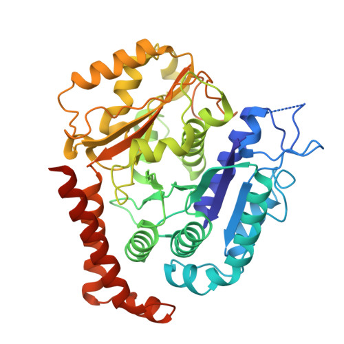
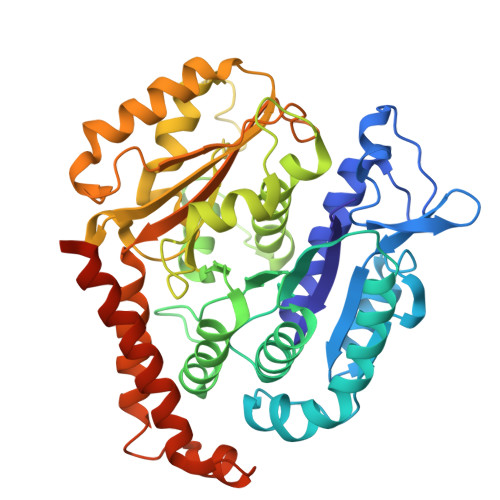
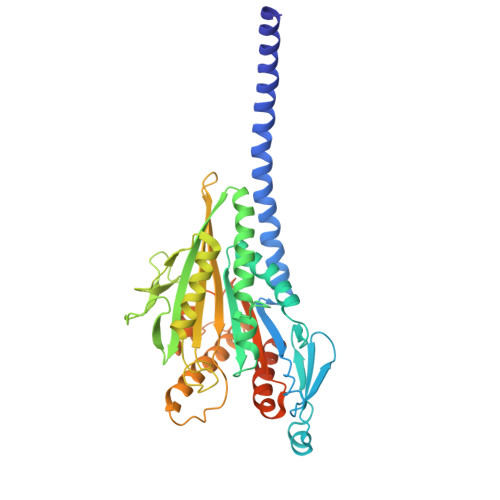
8YY2 | pdb_00008yy2

Kinesin-14 in nucleotide-free state bound to 13 PF Microtubule


Experimental Data Snapshot

  • Method: ELECTRON MICROSCOPY
  • Resolution: 3.60 Å
  • Aggregation State: FILAMENT 
  • Reconstruction Method: HELICAL 

wwPDB Validation   3D Report Full Report


This is version 2.0 of the entry. See complete history


Literature

Structural transitions in kinesin minus-end directed microtubule motility.

Shibata, S.Wang, M.Y.Imasaki, T.Shigematsu, H.Wei, Y.Jobichen, C.Hagio, H.Sivaraman, J.Endow, S.A.Nitta, R.

(2024) bioRxiv 

  • DOI: https://doi.org/10.1101/2024.07.29.605428
  • Primary Citation of Related Structures:  
    8YY2, 8YY3, 8YY4, 8YY5

  • PubMed Abstract: 

    Kinesin motor proteins hydrolyze ATP to produce force for spindle assembly and vesicle transport, performing essential functions in cell division and motility, but the structural changes required for force generation are uncertain. We now report high-resolution structures showing new transitions in the kinesin mechanochemical cycle, including power stroke fluctuations upon ATP binding and a post-hydrolysis state with bound ADP + free phosphate. We find that rate-limiting ADP release occurs upon microtubule binding, accompanied by central β-sheet twisting, which triggers the power stroke - stalk rotation and neck mimic docking - upon ATP binding. Microtubule release occurs with β-strand-to-loop transitions, implying that β-strand refolding induces Pi release and the recovery stroke. The strained β-sheet during the power stroke and strand-to-loop transitions identify the β-sheet as the long-sought motor spring.


  • Organizational Affiliation
    • Division of Structural Medicine and Anatomy, Department of Physiology and Cell Biology, Kobe University Graduate School of Medicine, Kobe, 650-0017, Japan.

Macromolecules
Find similar proteins by:  (by identity cutoff)  |  3D Structure
Entity ID: 1
MoleculeChains Sequence LengthOrganismDetailsImage
Tubulin alpha-1B chain451Sus scrofaMutation(s): 0 
EC: 3.6.5
UniProt
Find proteins for Q2XVP4 (Sus scrofa)
Explore Q2XVP4 
Go to UniProtKB:  Q2XVP4
Entity Groups  
Sequence Clusters30% Identity50% Identity70% Identity90% Identity95% Identity100% Identity
UniProt GroupQ2XVP4
Sequence Annotations
Expand
  • Reference Sequence
Find similar proteins by:  (by identity cutoff)  |  3D Structure
Entity ID: 2
MoleculeChains Sequence LengthOrganismDetailsImage
Tubulin beta chain444Sus scrofaMutation(s): 0 
UniProt
Find proteins for Q767L7 (Sus scrofa)
Explore Q767L7 
Go to UniProtKB:  Q767L7
Entity Groups  
Sequence Clusters30% Identity50% Identity70% Identity90% Identity95% Identity100% Identity
UniProt GroupQ767L7
Sequence Annotations
Expand
  • Reference Sequence
Find similar proteins by:  (by identity cutoff)  |  3D Structure
Entity ID: 3
MoleculeChains Sequence LengthOrganismDetailsImage
Protein claret segregational
C, D
409Drosophila melanogasterMutation(s): 3 
Gene Names: ncdCA(ND)CG7831
EC: 3.6.4
UniProt
Find proteins for P20480 (Drosophila melanogaster)
Explore P20480 
Go to UniProtKB:  P20480
Entity Groups  
Sequence Clusters30% Identity50% Identity70% Identity90% Identity95% Identity100% Identity
UniProt GroupP20480
Sequence Annotations
Expand
  • Reference Sequence
Small Molecules
Ligands 4 Unique
IDChains Name / Formula / InChI Key2D Diagram3D Interactions
GTP (Subject of Investigation/LOI)
Query on GTP

Download Ideal Coordinates CCD File 
E [auth B]GUANOSINE-5'-TRIPHOSPHATE
C10 H16 N5 O14 P3
XKMLYUALXHKNFT-UUOKFMHZSA-N
GDP (Subject of Investigation/LOI)
Query on GDP

Download Ideal Coordinates CCD File 
F [auth B]GUANOSINE-5'-DIPHOSPHATE
C10 H15 N5 O11 P2
QGWNDRXFNXRZMB-UUOKFMHZSA-N
ADP (Subject of Investigation/LOI)
Query on ADP

Download Ideal Coordinates CCD File 
H [auth D]ADENOSINE-5'-DIPHOSPHATE
C10 H15 N5 O10 P2
XTWYTFMLZFPYCI-KQYNXXCUSA-N
MG (Subject of Investigation/LOI)
Query on MG

Download Ideal Coordinates CCD File 
G [auth D]MAGNESIUM ION
Mg
JLVVSXFLKOJNIY-UHFFFAOYSA-N
Experimental Data & Validation

Experimental Data

  • Method: ELECTRON MICROSCOPY
  • Resolution: 3.60 Å
  • Aggregation State: FILAMENT 
  • Reconstruction Method: HELICAL 
EM Software:
TaskSoftware PackageVersion
MODEL REFINEMENTPHENIX

Structure Validation

View Full Validation Report


Entry History & Funding Information

Deposition Data


Funding OrganizationLocationGrant Number
Japan Science and TechnologyJapanJPMJFR214K
Japan Science and TechnologyJapanJPMJMS2024
Japan Agency for Medical Research and Development (AMED)JapanJP21gm1610003
Japan Society for the Promotion of Science (JSPS)Japan22K06809

Revision History  (Full details and data files)

  • Version 1.0: 2025-10-08
    Type: Initial release
  • Version 2.0: 2025-10-22
    Changes: Advisory, Atomic model, Author supporting evidence, Data collection, Derived calculations