8YUD | pdb_00008yud

Crystal structure of Xylose isomerase from Streptomyces avermitilis


Experimental Data Snapshot

  • Method: X-RAY DIFFRACTION
  • Resolution: 2.81 Å
  • R-Value Free: 
    0.243 (Depositor), 0.240 (DCC) 
  • R-Value Work: 
    0.187 (Depositor), 0.190 (DCC) 
  • R-Value Observed: 
    0.189 (Depositor) 

Starting Model: experimental
View more details

wwPDB Validation   3D Report Full Report


This is version 1.1 of the entry. See complete history


Literature

Data of the crystal structure of xylose isomerase from Streptomyces avermitilis.

Nam, K.H.

(2025) Data Brief 59: 111414-111414

  • DOI: https://doi.org/10.1016/j.dib.2025.111414
  • Primary Citation of Related Structures:  
    8YUD

  • PubMed Abstract: 

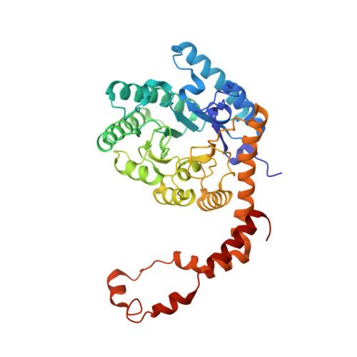
    Xylose isomerase (XI; also known as glucose isomerase) catalyzes the conversion of D-glucose and D-xylose to D-fructose and D-xylulose, respectively. XI is widely used in various industries, such as high-fructose corn syrup and bioethanol production. The discovery and characterization of novel XI variants are important to enhance the effective industrial applications of XI. Recently, the X-ray diffraction data for XI from Streptomyces avermitilis (SavXI) were collected at a synchrotron. The crystal structure of SavXI was determined using the molecular replacement method. The SavXI structure exhibited two unique metal-binding sites at the active site, diverse conformations, and a distinctive conformation of the C-terminal region compared to other XI homologs. This structural information extends the understanding of the molecular properties of the XI family. Here, information on the raw diffraction data images of SavXI, which were not presented in the previous study, is introduced. Detailed data collection and structure determination are reported for future XI structural analyses.


  • Organizational Affiliation
    • College of General Education, Kookmin University, Seoul 02707, Republic of Korea.

Macromolecules
Find similar proteins by:  (by identity cutoff)  |  3D Structure
Entity ID: 1
MoleculeChains Sequence LengthOrganismDetailsImage
Xylose isomerase
A, B, C, D, E
A, B, C, D, E, F
388Streptomyces avermitilisMutation(s): 0 
Gene Names: xylASAV14893_068640SAV31267_017200
EC: 5.3.1.5
UniProt
Find proteins for Q93HF3 (Streptomyces avermitilis (strain ATCC 31267 / DSM 46492 / JCM 5070 / NBRC 14893 / NCIMB 12804 / NRRL 8165 / MA-4680))
Explore Q93HF3 
Go to UniProtKB:  Q93HF3
Entity Groups  
Sequence Clusters30% Identity50% Identity70% Identity90% Identity95% Identity100% Identity
UniProt GroupQ93HF3
Sequence Annotations
Expand
  • Reference Sequence
Small Molecules
Ligands 1 Unique
IDChains Name / Formula / InChI Key2D Diagram3D Interactions
MG (Subject of Investigation/LOI)
Query on MG

Download Ideal Coordinates CCD File 
G [auth A]
H [auth A]
I [auth B]
J [auth B]
K [auth C]
G [auth A],
H [auth A],
I [auth B],
J [auth B],
K [auth C],
L [auth C],
M [auth D],
N [auth D],
O [auth E],
P [auth E],
Q [auth F],
R [auth F]
MAGNESIUM ION
Mg
JLVVSXFLKOJNIY-UHFFFAOYSA-N
Experimental Data & Validation

Experimental Data

  • Method: X-RAY DIFFRACTION
  • Resolution: 2.81 Å
  • R-Value Free:  0.243 (Depositor), 0.240 (DCC) 
  • R-Value Work:  0.187 (Depositor), 0.190 (DCC) 
  • R-Value Observed: 0.189 (Depositor) 
Space Group: P 32 2 1
Unit Cell:
Length ( Å )Angle ( ˚ )
a = 129.135α = 90
b = 129.135β = 90
c = 233.058γ = 120
Software Package:
Software NamePurpose
PHENIXrefinement
HKL-2000data reduction
HKL-2000data scaling
MOLREPphasing

Structure Validation

View Full Validation Report


Entry History & Funding Information

Deposition Data

  • Released Date: 2024-04-10 
  • Deposition Author(s): Nam, K.H.

Funding OrganizationLocationGrant Number
National Research Foundation (NRF, Korea)Korea, Republic OfNRF-2021R1I1A1A01050838

Revision History  (Full details and data files)

  • Version 1.0: 2024-04-10
    Type: Initial release
  • Version 1.1: 2025-06-25
    Changes: Database references, Structure summary