alpha-Amino-beta-carboxymuconate-epsilon-semialdehyde decarboxylase catalyzes enol/keto tautomerization of oxaloacetate.
Yang, Y., Davis, I., Altman, R.A., Liu, A.(2024) J Biol Chem 300: 107878-107878
- PubMed: 39395800 Search on PubMedSearch on PubMed Central
- DOI: https://doi.org/10.1016/j.jbc.2024.107878
- Primary Citation Related Structures:
8YT1, 8YT2 - PubMed Abstract:
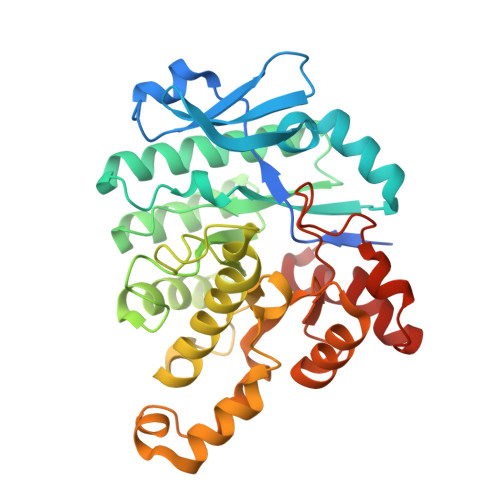
ACMSD (α-amino-β-carboxymuconate-ε-semialdehyde decarboxylase) is a key metalloenzyme critical for regulating de novo endogenous NAD + /NADH biosynthesis through the tryptophan-kynurenine pathway. This decarboxylase is a recognized target implicated in mitochondrial diseases and neurodegenerative disorders. However, unraveling its enzyme-substrate complex has been challenging due to its high catalytic efficiency. Here, we present a combined biochemical and structural study wherein we determined the crystal structure of ACMSD in complex with malonate. Our analysis revealed significant rearrangements in the active site, particularly in residues crucial for ACMS decarboxylation, including Arg51, Arg239∗ (a residue from an adjacent subunit), His228, and Trp194. Docking modeling studies proposed a putative ACMS binding mode. Additionally, we found that ACMSD catalyzes oxaloacetic acid (OAA) tautomerization at a rate of 6.51 ± 0.42 s -1 but not decarboxylation. The isomerase activity of ACMSD on OAA warrants further investigation in future biological studies. Subsequent mutagenesis studies and crystallographic analysis of the W194A variant shed light on the roles of specific second-coordination sphere residues. Our findings indicate that Arg51 and Arg239∗ are crucial for OAA tautomerization. Moreover, our comparative analysis with related isomerase superfamily members underscores a general strategy employing arginine residues to promote OAA isomerization. Given the observed isomerase activity of ACMSD on OAA and its structural similarity to ACMS, we propose that ACMSD may facilitate isomerization to ensure ACMS is in the optimal tautomeric form for subsequent decarboxylation initiated by the zinc-bound hydroxide ion. Overall, these findings deepen the understanding of the structure and function of ACMSD, offering insights into potential therapeutic interventions.
- State Key Laboratory of Biocatalysis and Enzyme Engineering, Hubei Collaborative Innovation Center for Green Transformation of Bio-Resources, Hubei Key Laboratory of Industrial Biotechnology, School of Life Sciences, Hubei University, Wuhan, China; Department of Chemistry, University of Texas at San Antonio, San Antonio, Texas, USA. Electronic address: yangyu@hubu.edu.cn.
Organizational Affiliation: