8YSB | pdb_00008ysb

Crystal structure of DynA1, a putative monoxygenase from Mivromonospora chersina.


Experimental Data Snapshot

  • Method: X-RAY DIFFRACTION
  • Resolution: 2.30 Å
  • R-Value Free: 
    0.289 (Depositor), 0.292 (DCC) 
  • R-Value Work: 
    0.266 (Depositor), 0.269 (DCC) 
  • R-Value Observed: 
    0.268 (Depositor) 

Starting Model: in silico
View more details

wwPDB Validation 3D Report Full Report

Validation slider image for 8YSB

This is version 1.1 of the entry. See complete history

Literature

An Enzymatic Oxidation Cascade Converts delta-Thiolactone Anthracene to Anthraquinone in the Biosynthesis of Anthraquinone-Fused Enediynes.

Ma, G.L.Liu, W.Q.Huang, H.Yan, X.F.Shen, W.Visitsatthawong, S.Prakinee, K.Tran, H.Fan, X.Gao, Y.G.Chaiyen, P.Li, J.Liang, Z.X.

(2024) JACS Au 4: 2925-2935

  • DOI: https://doi.org/10.1021/jacsau.4c00279
  • Primary Citation Related Structures: 
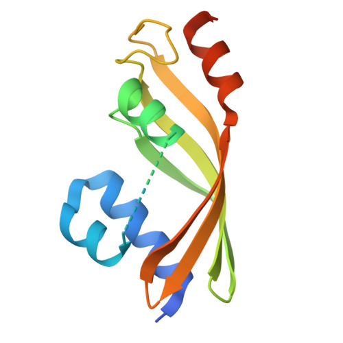
    8YSB

  • PubMed Abstract: 

    Anthraquinone-fused enediynes are anticancer natural products featuring a DNA-intercalating anthraquinone moiety. Despite recent insights into anthraquinone-fused enediyne (AQE) biosynthesis, the enzymatic steps involved in anthraquinone biogenesis remain to be elucidated. Through a combination of in vitro and in vivo studies, we demonstrated that a two-enzyme system, composed of a flavin adenine dinucleotide (FAD)-dependent monooxygenase (DynE13) and a cofactor-free enzyme (DynA1), catalyzes the final steps of anthraquinone formation by converting δ-thiolactone anthracene to hydroxyanthraquinone. We showed that the three oxygen atoms in the hydroxyanthraquinone originate from molecular oxygen (O 2 ), with the sulfur atom eliminated as H 2 S. We further identified the key catalytic residues of DynE13 and A1 by structural and site-directed mutagenesis studies. Our data support a catalytic mechanism wherein DynE13 installs two oxygen atoms with concurrent desulfurization and decarboxylation, whereas DynA1 acts as a cofactor-free monooxygenase, installing the final oxygen atom in the hydroxyanthraquinone. These findings establish the indispensable roles of DynE13 and DynA1 in AQE biosynthesis and unveil novel enzymatic strategies for anthraquinone formation.


  • Organizational Affiliation
    • School of Biological Sciences, Nanyang Technological University, Singapore 637551, Singapore.

Macromolecule Content 

  • Total Structure Weight: 15.71 kDa 
  • Atom Count: 835 
  • Modeled Residue Count: 110 
  • Deposited Residue Count: 146 
  • Unique protein chains: 1

Macromolecules

Find similar proteins by:|  3D Structure
Entity ID: 1
MoleculeChains  Sequence LengthOrganismDetailsImage
Predicted ester cyclase146Micromonospora chersinaMutation(s): 1 
Gene Names: GA0070603_4190
UniProt
Find proteins for B2BM50 (Micromonospora chersina)
Explore B2BM50 
Go to UniProtKB:  B2BM50
Entity Groups
Sequence Clusters30% Identity50% Identity70% Identity90% Identity95% Identity100% Identity
UniProt GroupB2BM50
Sequence Annotations
Expand
Reference Sequence

Experimental Data & Validation

Experimental Data

  • Method: X-RAY DIFFRACTION
  • Resolution: 2.30 Å
  • R-Value Free:  0.289 (Depositor), 0.292 (DCC) 
  • R-Value Work:  0.266 (Depositor), 0.269 (DCC) 
  • R-Value Observed: 0.268 (Depositor) 
Space Group: C 2 2 21
Unit Cell:
Length ( Å )Angle ( ˚ )
a = 33.976α = 90
b = 84.16β = 90
c = 83.228γ = 90
Software Package:
Software NamePurpose
PHENIXrefinement
XDSdata reduction
XDSdata scaling
PHASERphasing
Cootmodel building

Structure Validation

View Full Validation Report


Entry History 

& Funding Information

Deposition Data


Funding OrganizationLocationGrant Number
Ministry of Education (MoE, Singapore)SingaporeMOE2019-T2-2-099
Ministry of Education (MoE, Singapore)SingaporeRG108/20

Revision History  (Full details and data files)

  • Version 1.0: 2024-09-04
    Type: Initial release
  • Version 1.1: 2024-09-11
    Changes: Database references