N-terminal helix formation and dimer-monomer transition of FGF10 in specific recognition of FGFR2b.
Park, H., Park, Y.S., Kwak, K., Yun, J., Lee, H., Lee, C.E., Lee, D., Lee, K.W., An, Y.J., Yim, H.S., Lee, J.H., Cha, S.S., Kang, L.W.(2025) FEBS J 292: 6735-6746
- PubMed: 40794773 Search on PubMed
- DOI: https://doi.org/10.1111/febs.70218
- Primary Citation Related Structures:
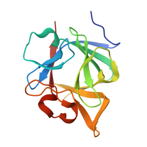
8YQ1 - PubMed Abstract:
Fibroblast growth factors (FGFs) are crucial for various cellular functions, including proliferation, differentiation, tissue repair, and immune responses. FGF10, part of the FGF7 subfamily, binds to the FGFR2b receptor via heparin sulfate. We determined the crystal structure of human FGF10 alone. In the FGFR2b-bound form, the N-terminal region of FGF10 formed an α1 helix. This α1 helix, however, exists as a flexible loop with dual conformations in the unbound structure. Deleting this conformationally dynamic α1 helix reduces cell proliferation activity in vitro. Receptor-binding-induced formation of the α1 helix in FGF10 creates a distinct protruded knob and concave pocket on the globular core of FGFs, increasing the FGFR2b-binding surface by 37%. Size-exclusion chromatography showed a concentration-dependent dimer-monomer shift in purified FGF10, with the hydrophobic dimer interface aligning with the FGFR2b D2-domain-binding surface. These findings suggest that the conformational change in the N-terminal region and the dimer-monomer shift are critical for FGF10's specific binding to FGFR2b, highlighting the functional significance of these structural adaptations.
- Department of Biological Sciences, Konkuk University, Seoul, Republic of Korea.
Organizational Affiliation: