Mechanism and Utility of the ATP-Grasp Enzyme BesA for the Synthesis of Non-natural Alkyne-Containing Dipeptides Applicable for Click Chemistry.
Otsuka, H., Fujishiro, T.(2025) ACS Chem Biol 20: 2521-2532
- PubMed: 41047544 Search on PubMed
- DOI: https://doi.org/10.1021/acschembio.5c00676
- Primary Citation Related Structures:
8YI6 - PubMed Abstract:
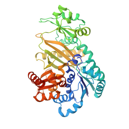
Terminal alkyne-containing biomolecules are key compounds utilized in bioorthogonal chemistry via azide-alkyne cycloaddition click chemistry. Various synthetic strategies for the introduction of the terminal alkyne to biomolecules have been developed; however, an enzymatic terminal alkyne-modifying system is not well-explored because the biosynthetic systems for terminal alkynes are rare. Recently, BesA, a member of the ATP-grasp enzyme family, has been reported to exclusively utilize terminal alkyne-containing l-propargylglycine and l-glutamic acid as substrates in the synthesis of γ-l-glutamyl-l-propargylglycine. Because of its use of the terminal alkyne for click chemistry, a BesA-based catalytic system is regarded as a potentially attractive biocatalyst for the enrichment of terminal alkyne-containing biomolecules. Toward developing BesA-based biocatalysts, it is important to understand the structure-based mechanism of action of BesA, especially recognition of the terminal alkyne. Here, we elucidate the structural basis of BesA for synthesis of γ-l-glutamyl-l-propargylglycine. The X-ray crystal analysis of BesA unveiled a narrow substrate-binding cleft, beside Y33, R50, R365, and R404 as conserved residues among BesA enzymes from Streptomyces , as the active site for binding of two amino acids, l-propargylglycine and l-glutamic acid. In particular, the region beside Y33 is likely to accommodate the terminal alkyne of l-propargylglycine via CH-π interaction based on the dipeptide-docking simulation of BesA and the results of the activity assay of the BesA Y33A variant. Furthermore, we demonstrate a BesA-catalyzed conjugation system for the synthesis of non-natural alkyne-containing dipeptides. The BesA R50A variant showed a little activity for ligation between l-propargylglycine and 1-methyl-l-glutamate, affording 1-methyl-l-glutamyl-l-propargylglycine. Moreover, the BesA wild-type showed activity for ligation of l-homopropargylglycine and l-glutamic acid, yielding γ-l-glutamyl-l-homopropargylglycine. Structural comparison of BesA with proteins that possibly bind the alkynes shows the significance of Tyr in recognition of the alkynes. These findings highlight the usefulness of BesA-based biocatalytic systems in expanding the chemical space of alkyne-containing peptides applicable for click chemistry as well as understanding alkyne recognition by proteins.
- Department of Biochemistry and Molecular Biology, Graduate School of Science and Engineering, Saitama University, Shimo-okubo 255, Sakura-ku, Saitama 338-8570Japan.
Organizational Affiliation: