Cooperation of a polymerizing SAM domain and an intrinsically disordered region enables full SAMD1 function on chromatin.
Geller, M., Cao, Y., Simon, C., Stielow, B., Xu, J., Wei, P., Nist, A., Rohner, I., Jeude, L.M., Huber, T., Stiewe, T., Wang, Z., Liefke, R.(2025) Nucleic Acids Res 53
- PubMed: 40183636 Search on PubMedSearch on PubMed Central
- DOI: https://doi.org/10.1093/nar/gkaf259
- Primary Citation Related Structures:
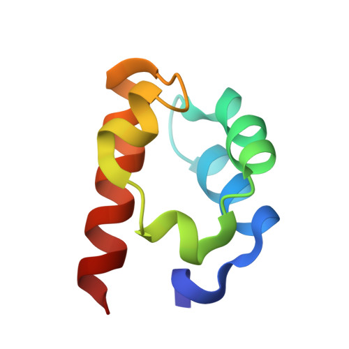
8Y76, 8Y77 - PubMed Abstract:
Transcription factors orchestrate gene expression through a myriad of complex mechanisms, encompassing collaborations with other transcription factors and the formation of multimeric complexes. The chromatin-binding protein SAMD1 [sterile alpha motif (SAM) domain-containing protein 1] binds to unmethylated CpG-rich DNA utilizing its N-terminal winged-helix (WH) domain. Additionally, its C-terminal SAM domain, which mediates interactions with itself and with L3MBTL3, is crucial for chromatin binding. The precise role of the SAM domain in this process remains unclear. Using structural analyses, we elucidated the distinct homopolymerization modes within the SAM domains of L3MBTL3 and SAMD1, alongside their heterodimerization architecture. Interestingly, SAMD1 necessitates not only the WH and SAM domain but also a proline/alanine-rich intrinsically disordered region (IDR) for efficient chromatin binding. The IDR is essential for the ability of SAMD1 to form large polymers, with its functionality determined by integrity rather than the specific sequence. Mutagenesis studies underscore the critical role of arginines within the IDR for polymerization, chromatin binding, and the biological function of SAMD1. These findings propose a model in which structured and unstructured regions of SAMD1 cooperate in a coordinated fashion to facilitate chromatin binding. This work provides new insights into the diverse mechanisms transcription factors employ to interact with chromatin and regulate gene expression.
- Institute of Molecular Biology and Tumor Research (IMT), Philipps University of Marburg, Marburg 35043, Germany.
Organizational Affiliation: