


The Crystal Structure of Apaf from Biortus.
Wang, F., Cheng, W., Lv, Z., Ju, C., Ni, C.To be published.
Experimental Data Snapshot
Starting Model: experimental
View more details
Entity ID: 1 | |||||
|---|---|---|---|---|---|
| Molecule | Chains | Sequence Length | Organism | Details | Image |
| Apoptotic protease-activating factor 1 | 98 | Homo sapiens | Mutation(s): 0 Gene Names: APAF1, KIAA0413 | 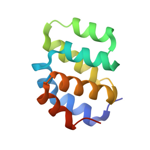 | |
UniProt & NIH Common Fund Data Resources | |||||
PHAROS: O14727 GTEx: ENSG00000120868 | |||||
Entity Groups | |||||
| Sequence Clusters | 30% Identity50% Identity70% Identity90% Identity95% Identity100% Identity | ||||
| UniProt Group | O14727 | ||||
Sequence AnnotationsExpand | |||||
Reference Sequence | |||||
| Ligands 1 Unique | |||||
|---|---|---|---|---|---|
| ID | Chains | Name / Formula / InChI Key | 2D Diagram | 3D Interactions | |
| PO4 (Subject of Investigation/LOI) Download:Ideal Coordinates CCD File | E [auth A], F [auth B], G [auth B], H [auth C], I [auth D] | PHOSPHATE ION O4 P NBIIXXVUZAFLBC-UHFFFAOYSA-K |  | ||
| Length ( Å ) | Angle ( ˚ ) |
|---|---|
| a = 31.641 | α = 90 |
| b = 75.336 | β = 90 |
| c = 161.067 | γ = 90 |
| Software Name | Purpose |
|---|---|
| REFMAC | refinement |
| XDS | data reduction |
| Aimless | data scaling |
| PHASER | phasing |


