8XMK | pdb_00008xmk

Local refinement of SRCR5-9 domains

  • Classification: ENDOCYTOSIS
  • Organism(s): Homo sapiens
  • Expression System: Homo sapiens
  • Mutation(s): No 

  • Deposited: 2023-12-27 Released: 2025-01-01 
  • Deposition Author(s): Xu, H., Su, X.D.
  • Funding Organization(s): Ministry of Science and Technology (MoST, China)

Experimental Data Snapshot

  • Method: ELECTRON MICROSCOPY
  • Resolution: 3.03 Å
  • Aggregation State: PARTICLE 
  • Reconstruction Method: SINGLE PARTICLE 

wwPDB Validation   3D Report Full Report


This is version 1.2 of the entry. See complete history


Literature

Calcium-dependent oligomerization of scavenger receptor CD163 facilitates the endocytosis of ligands.

Xu, H.Song, X.Su, X.D.

(2025) Nat Commun 16: 6679-6679

  • DOI: https://doi.org/10.1038/s41467-025-62013-4
  • Primary Citation of Related Structures:  
    8XMK, 8XMP, 8XMQ, 8XMW

  • PubMed Abstract: 

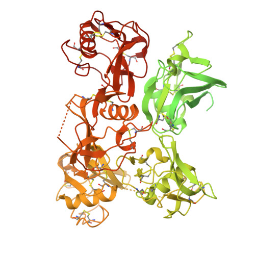
    Scavenger receptor CD163 is a marker of M2 type macrophages that play important roles in anti-inflammatory processes. The most extensively studied function of CD163 is related to the elimination of hemoglobin-haptoglobin (Hb-Hp) complexes, to prevent potential oxidative toxicity of the iron-containing heme. However, the structural mechanism of CD163 in ligand binding and internalization remains elusive. Here, we present the cryo-electron microscopy structure of human Hb-Hp recognition by the full ectodomain of CD163. We illuminate that CD163 forms calcium-dependent oligomers and primarily exists as trimeric form under the condition of 2.5 mM calcium. It mainly utilizes two protomers to interact with Hb-Hp complex asymmetrically, while the third protomer of the trimer also has the potential to form calcium-mediated contacts with Hp. Flow cytometry analyses reveal that oligomerization of CD163 significantly enhances the efficiency of ligand endocytosis. These results advance our understanding of the role of CD163 in ligand scavenging.


  • Organizational Affiliation
    • Biomedical Pioneering Innovation Center (BIOPIC), and State Key Laboratory of Gene Function and Modulation Research, School of Life Sciences, Peking University, Beijing, China. xuhua@pku.edu.cn.

Macromolecules
Find similar proteins by:  (by identity cutoff)  |  3D Structure
Entity ID: 1
MoleculeChains Sequence LengthOrganismDetailsImage
Scavenger receptor cysteine-rich type 1 protein M130A [auth D],
B [auth E],
C [auth F]
1,009Homo sapiensMutation(s): 0 
Gene Names: CD163M130
UniProt & NIH Common Fund Data Resources
Find proteins for Q86VB7 (Homo sapiens)
Explore Q86VB7 
Go to UniProtKB:  Q86VB7
PHAROS:  Q86VB7
GTEx:  ENSG00000177575 
Entity Groups  
Sequence Clusters30% Identity50% Identity70% Identity90% Identity95% Identity100% Identity
UniProt GroupQ86VB7
Glycosylation
Glycosylation Sites: 2Go to GlyGen: Q86VB7-1
Sequence Annotations
Expand
  • Reference Sequence
Small Molecules
Ligands 2 Unique
IDChains Name / Formula / InChI Key2D Diagram3D Interactions
NAG
Query on NAG

Download Ideal Coordinates CCD File 
D
E [auth D]
L [auth E]
M [auth E]
S [auth F]
D,
E [auth D],
L [auth E],
M [auth E],
S [auth F],
T [auth F]
2-acetamido-2-deoxy-beta-D-glucopyranose
C8 H15 N O6
OVRNDRQMDRJTHS-FMDGEEDCSA-N
CA
Query on CA

Download Ideal Coordinates CCD File 
F [auth D]
G [auth D]
H [auth D]
I [auth D]
J [auth D]
F [auth D],
G [auth D],
H [auth D],
I [auth D],
J [auth D],
K [auth D],
N [auth E],
O [auth E],
P [auth E],
Q [auth E],
R [auth E],
U [auth F],
V [auth F],
W [auth F],
X [auth F],
Y [auth F],
Z [auth F]
CALCIUM ION
Ca
BHPQYMZQTOCNFJ-UHFFFAOYSA-N
Experimental Data & Validation

Experimental Data

  • Method: ELECTRON MICROSCOPY
  • Resolution: 3.03 Å
  • Aggregation State: PARTICLE 
  • Reconstruction Method: SINGLE PARTICLE 
EM Software:
TaskSoftware PackageVersion
MODEL REFINEMENTPHENIX1.20.1_4487:

Structure Validation

View Full Validation Report


Entry History & Funding Information

Deposition Data


Funding OrganizationLocationGrant Number
Ministry of Science and Technology (MoST, China)China2021YFC2301400

Revision History  (Full details and data files)

  • Version 1.0: 2025-01-01
    Type: Initial release
  • Version 1.1: 2025-07-30
    Changes: Data collection, Database references
  • Version 1.2: 2025-08-06
    Changes: Data collection, Database references